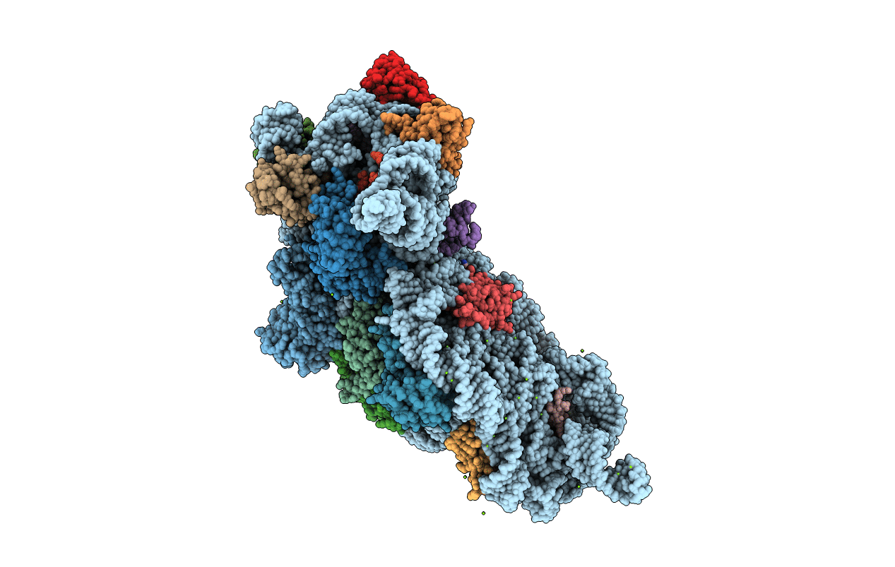
6CAR image
Entry Detail
PDB ID:
6CAR
Keywords:
Title:
Serial Femtosecond X-ray Crystal Structure of 30S ribosomal subunit from Thermus thermophilus in complex with Sisomicin
Biological Source:
Source Organism:
PDB Version:
Deposition Date:
2018-01-31
Release Date:
2018-07-25
Method Details:
Experimental Method:
Resolution:
3.40 Å
R-Value Free:
0.26
R-Value Work:
0.22
R-Value Observed:
0.22
Space Group:
P 41 21 2
Macromolecular Entities
Polymer Type:polyribonucleotide
Description:16S Ribosomal RNA rRNA
Chain IDs:A
Chain Length:1517
Number of Molecules:1
Biological Source:Thermus thermophilus HB8
Polymer Type:polypeptide(L)
Description:30S ribosomal protein S2
Chain IDs:B
Chain Length:255
Number of Molecules:1
Biological Source:Thermus thermophilus (strain HB8 / ATCC 27634 / DSM 579)
Polymer Type:polypeptide(L)
Description:30S ribosomal protein S3
Chain IDs:C
Chain Length:238
Number of Molecules:1
Biological Source:Thermus thermophilus (strain HB8 / ATCC 27634 / DSM 579)
Polymer Type:polypeptide(L)
Description:30S ribosomal protein S4
Chain IDs:D
Chain Length:208
Number of Molecules:1
Biological Source:Thermus thermophilus (strain HB8 / ATCC 27634 / DSM 579)
Polymer Type:polypeptide(L)
Description:30S ribosomal protein S5
Chain IDs:E
Chain Length:161
Number of Molecules:1
Biological Source:Thermus thermophilus (strain HB8 / ATCC 27634 / DSM 579)
Polymer Type:polypeptide(L)
Description:30S ribosomal protein S6
Chain IDs:F
Chain Length:101
Number of Molecules:1
Biological Source:Thermus thermophilus (strain HB8 / ATCC 27634 / DSM 579)
Polymer Type:polypeptide(L)
Description:30S ribosomal protein S7
Chain IDs:G
Chain Length:155
Number of Molecules:1
Biological Source:Thermus thermophilus (strain HB8 / ATCC 27634 / DSM 579)
Polymer Type:polypeptide(L)
Description:30S ribosomal protein S8
Chain IDs:H
Chain Length:138
Number of Molecules:1
Biological Source:Thermus thermophilus (strain HB8 / ATCC 27634 / DSM 579)
Polymer Type:polypeptide(L)
Description:30S ribosomal protein S9
Chain IDs:I
Chain Length:127
Number of Molecules:1
Biological Source:Thermus thermophilus (strain HB8 / ATCC 27634 / DSM 579)
Polymer Type:polypeptide(L)
Description:30S ribosomal protein S10
Chain IDs:J
Chain Length:104
Number of Molecules:1
Biological Source:Thermus thermophilus (strain HB8 / ATCC 27634 / DSM 579)
Polymer Type:polypeptide(L)
Description:30S ribosomal protein S11
Chain IDs:K
Chain Length:128
Number of Molecules:1
Biological Source:Thermus thermophilus (strain HB8 / ATCC 27634 / DSM 579)
Polymer Type:polypeptide(L)
Description:30S ribosomal protein S12
Chain IDs:L
Chain Length:131
Number of Molecules:1
Biological Source:Thermus thermophilus (strain HB8 / ATCC 27634 / DSM 579)
Polymer Type:polypeptide(L)
Description:30S ribosomal protein S13
Chain IDs:M
Chain Length:125
Number of Molecules:1
Biological Source:Thermus thermophilus (strain HB8 / ATCC 27634 / DSM 579)
Polymer Type:polypeptide(L)
Description:30S ribosomal protein S14 type Z
Chain IDs:N
Chain Length:60
Number of Molecules:1
Biological Source:Thermus thermophilus (strain HB8 / ATCC 27634 / DSM 579)
Polymer Type:polypeptide(L)
Description:30S ribosomal protein S15
Chain IDs:O
Chain Length:88
Number of Molecules:1
Biological Source:Thermus thermophilus (strain HB8 / ATCC 27634 / DSM 579)
Polymer Type:polypeptide(L)
Description:30S ribosomal protein S16
Chain IDs:P
Chain Length:88
Number of Molecules:1
Biological Source:Thermus thermophilus (strain HB8 / ATCC 27634 / DSM 579)
Polymer Type:polypeptide(L)
Description:30S ribosomal protein S17
Chain IDs:Q
Chain Length:104
Number of Molecules:1
Biological Source:Thermus thermophilus (strain HB8 / ATCC 27634 / DSM 579)
Polymer Type:polypeptide(L)
Description:30S ribosomal protein S18
Chain IDs:R
Chain Length:87
Number of Molecules:1
Biological Source:Thermus thermophilus (strain HB8 / ATCC 27634 / DSM 579)
Polymer Type:polypeptide(L)
Description:30S ribosomal protein S19
Chain IDs:S
Chain Length:92
Number of Molecules:1
Biological Source:Thermus thermophilus (strain HB8 / ATCC 27634 / DSM 579)
Polymer Type:polypeptide(L)
Description:30S ribosomal protein S20
Chain IDs:T
Chain Length:105
Number of Molecules:1
Biological Source:Thermus thermophilus (strain HB8 / ATCC 27634 / DSM 579)
Polymer Type:polypeptide(L)
Description:30S ribosomal protein Thx
Chain IDs:U
Chain Length:26
Number of Molecules:1
Biological Source:Thermus thermophilus (strain HB8 / ATCC 27634 / DSM 579)
Polymer Type:polyribonucleotide
Description:RNA (5'-R(*GP*GP*GP*AP*UP*UP*GP*AP*AP*AP*AP*UP*CP*CP*C)-3')
Chain IDs:W
Chain Length:15
Number of Molecules:1
Biological Source:Thermus thermophilus HB8
Polymer Type:polyribonucleotide
Description:RNA (5'-R(*UP*UP*UP*UP*UP*U)-3')
Chain IDs:V (auth: Y)
Chain Length:6
Number of Molecules:1
Biological Source:Thermus thermophilus HB8
Modified Residue
Compound ID Chain ID Parent Comp ID Details 2D Image
0TD L ASP modified residue
2MG A G modified residue
4OC A C modified residue
5MC A C modified residue
G7M A G modified residue
M2G A G modified residue
MA6 A A modified residue
PSU A U modified residue
UR3 A U modified residue
Primary Citation
Aminoglycoside ribosome interactions reveal novel conformational states at ambient temperature.
Nucleic Acids Res. 46 9793 9804 (2018)
PMID: 30113694 DOI: 10.1093/nar/gky693

Abstact

The bacterial 30S ribosomal subunit is a primary antibiotic target. Despite decades of discovery, the mechanisms by which antibiotic binding induces ribosomal dysfunction are not fully understood. Ambient temperature crystallographic techniques allow more biologically relevant investigation of how local antibiotic binding site interactions trigger global subunit rearrangements that perturb protein synthesis. Here, the structural effects of 2-deoxystreptamine (paromomycin and sisomicin), a novel sisomicin derivative, N1-methyl sulfonyl sisomicin (N1MS) and the non-deoxystreptamine (streptomycin) aminoglycosides on the ribosome at ambient and cryogenic temperatures were examined. Comparative studies led to three main observations. First, individual aminoglycoside-ribosome interactions in the decoding center were similar for cryogenic versus ambient temperature structures. Second, analysis of a highly conserved GGAA tetraloop of h45 revealed aminoglycoside-specific conformational changes, which are affected by temperature only for N1MS. We report the h44-h45 interface in varying states, i.e. engaged, disengaged and in equilibrium. Third, we observe aminoglycoside-induced effects on 30S domain closure, including a novel intermediary closure state, which is also sensitive to temperature. Analysis of three ambient and five cryogenic crystallography datasets reveal a correlation between h44-h45 engagement and domain closure. These observations illustrate the role of ambient temperature crystallography in identifying dynamic mechanisms of ribosomal dysfunction induced by local drug-binding site interactions. Together, these data identify tertiary ribosomal structural changes induced by aminoglycoside binding that provides functional insight and targets for drug design.

Legend

Protein

Chemical

Disease

Primary Citation of related structures