6C9H image
Deposition Date 2018-01-26
Release Date 2018-11-28
Last Version Date 2023-10-04
Entry Detail
PDB ID:
6C9H
Keywords:
Title:
non-phosphorylated AMP-activated protein kinase bound to pharmacological activator R734
Biological Source:
Source Organism:
Homo sapiens (Taxon ID: 9606)
Host Organism:
Method Details:
Experimental Method:
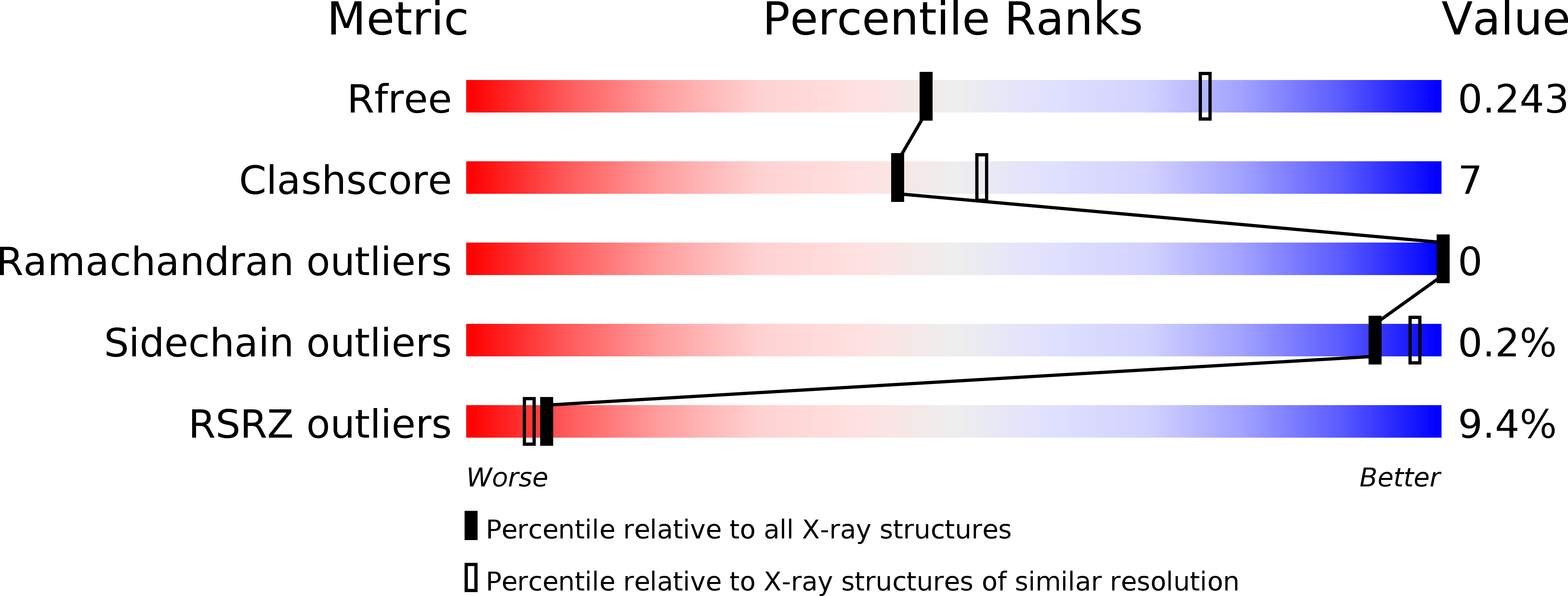
Resolution:
2.65 Å
R-Value Free:
0.24
R-Value Work:
0.21
R-Value Observed:
0.21
Space Group:
P 61 2 2
Macromolecular Entities
Polymer Type:polypeptide(L)
Molecule:5'-AMP-activated protein kinase catalytic subunit alpha-1
Gene (Uniprot):PRKAA1
Mutations:S108D
Chain IDs:A
Chain Length:494
Number of Molecules:1
Biological Source:Homo sapiens
Polymer Type:polypeptide(L)
Molecule:5'-AMP-activated protein kinase subunit beta-1
Gene (Uniprot):PRKAB1
Chain IDs:B
Chain Length:204
Number of Molecules:1
Biological Source:Homo sapiens
Polymer Type:polypeptide(L)
Molecule:5'-AMP-activated protein kinase subunit gamma-1
Gene (Uniprot):PRKAG1
Chain IDs:C
Chain Length:331
Number of Molecules:1
Biological Source:Homo sapiens
Primary Citation
Structures of AMP-activated protein kinase bound to novel pharmacological activators in phosphorylated, non-phosphorylated, and nucleotide-free states.
J. Biol. Chem. 294 953 967 (2019)
PMID: 30478170 DOI: 10.1074/jbc.RA118.004883

Abstact

AMP-activated protein kinase (AMPK) is an attractive therapeutic target for managing metabolic diseases. A class of pharmacological activators, including Merck 991, binds the AMPK ADaM site, which forms the interaction surface between the kinase domain (KD) of the α-subunit and the carbohydrate-binding module (CBM) of the β-subunit. Here, we report the development of two new 991-derivative compounds, R734 and R739, which potently activate AMPK in a variety of cell types, including β2-specific skeletal muscle cells. Surprisingly, we found that they have only minor effects on direct kinase activity of the recombinant α1β2γ1 isoform yet robustly enhance protection against activation loop dephosphorylation. This mode of activation is reminiscent of that of ADP, which activates AMPK by binding to the nucleotide-binding sites in the γ-subunit, more than 60 Å away from the ADaM site. To understand the mechanisms of full and partial AMPK activation, we determined the crystal structures of fully active phosphorylated AMPK α1β1γ1 bound to AMP and R734/R739 as well as partially active nonphosphorylated AMPK bound to R734 and AMP and phosphorylated AMPK bound to R734 in the absence of added nucleotides at <3-Å resolution. These structures and associated analyses identified a novel conformational state of the AMPK autoinhibitory domain associated with partial kinase activity and provide new insights into phosphorylation-dependent activation loop stabilization in AMPK.

Legend

Protein

Chemical

Disease

Primary Citation of related structures