6C9B image
Deposition Date 2018-01-26
Release Date 2018-04-04
Last Version Date 2023-11-15
Entry Detail
PDB ID:
6C9B
Title:
The structure of MppP soaked with the products 4HKA and 2KA
Biological Source:
Source Organism:
Host Organism:
Method Details:
Experimental Method:
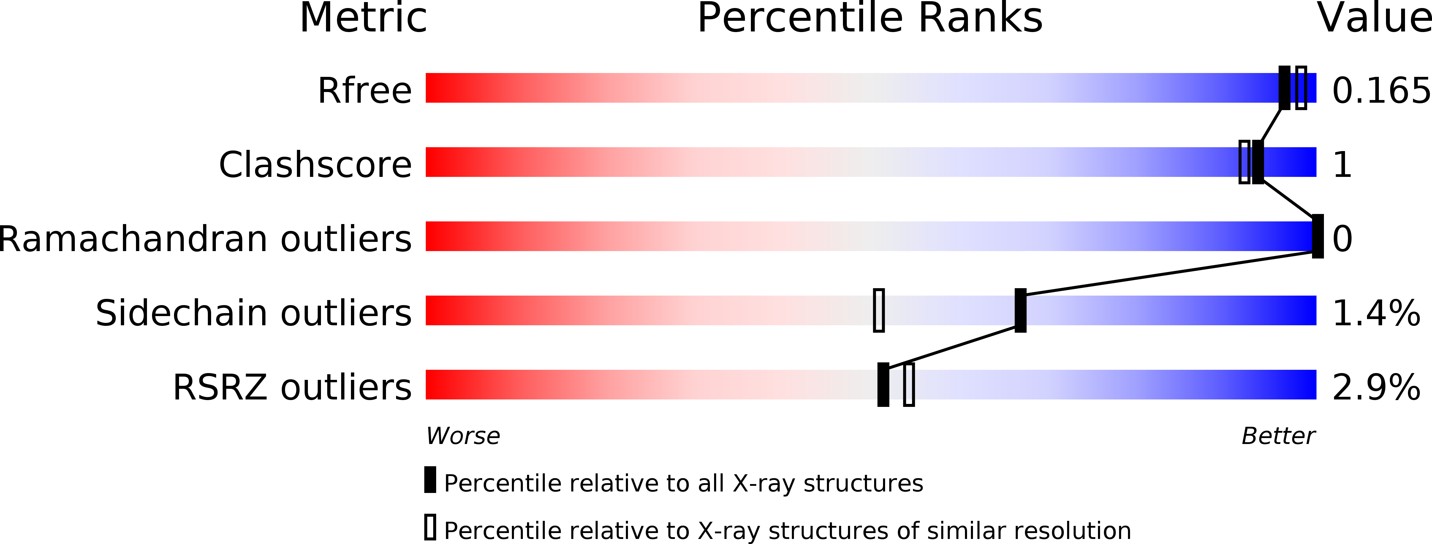
Resolution:
1.69 Å
R-Value Free:
0.16
R-Value Work:
0.15
R-Value Observed:
0.15
Space Group:
P 21 21 21
Macromolecular Entities
Polymer Type:polypeptide(L)
Molecule:PLP-Dependent L-Arginine Hydroxylase MppP
Chain IDs:A, B, C, D
Chain Length:376
Number of Molecules:4
Biological Source:Streptomyces wadayamensis
Modified Residue
Compound ID Chain ID Parent Comp ID Details 2D Image
LLP A LYS modified residue
Primary Citation
Streptomyces wadayamensis MppP is a PLP-Dependent Oxidase, Not an Oxygenase.
Biochemistry 57 3252 3264 (2018)
PMID: 29473729 DOI: 10.1021/acs.biochem.8b00130

Abstact

The PLP-dependent l-arginine hydroxylase/deaminase MppP from Streptomyces wadayamensis (SwMppP) is involved in the biosynthesis of l-enduracididine, a nonproteinogenic amino acid found in several nonribosomally produced peptide antibiotics. SwMppP uses only PLP and molecular oxygen to catalyze a 4-electron oxidation of l-arginine to form a mixture of 2-oxo-4(S)-hydroxy-5-guanidinovaleric acid and 2-oxo-5-guanidinovaleric acid. Steady-state kinetics analysis in the presence and absence of catalase shows that one molecule of peroxide is formed for every molecule of dioxygen consumed in the reaction. Moreover, for each molecule of 2-oxo-4(S)-hydroxy-5-guanidinovaleric acid produced, two molecules of dioxygen are consumed, suggesting that both the 4-hydroxy and 2-keto groups are derived from water. This was confirmed by running the reactions using either ([18])O(2) or H(2)([18])O and analyzing the products by ESI-MS. Incorporation of ([18])O was only observed when the reaction was performed in H(2)([18])O. Crystal structures of SwMppP with l-arginine, 2-oxo-4(S)-hydroxy-5-guanidinovaleric acid, or 2-oxo-5-guanidinovaleric acid bound were determined at resolutions of 2.2, 1.9. and 1.8 A, respectively. The structural data show that the N-terminal portion of the protein is disordered unless substrate or product is bound in the active site, in which case it forms a well-ordered helix that covers the catalytic center. This observation suggested that the N-terminal helix may have a role in substrate binding and/or catalysis. Our structural and kinetic characterizations of N-terminal variants show that the N-terminus is critical for catalysis. In light of this new information, we have refined our previously proposed mechanism of the SwMppP-catalyzed oxidation of l-arginine.

Legend

Protein

Chemical

Disease

Primary Citation of related structures