Deposition Date
2018-01-16
Release Date
2018-07-11
Last Version Date
2024-03-13
Entry Detail
PDB ID:
6C5R
Keywords:
Title:
Crystal structure of the soluble domain of the mitochondrial calcium uniporter
Biological Source:
Source Organism(s):
Metarhizium acridum (strain CQMa 102) (Taxon ID: 655827)
Expression System(s):
Method Details:
Experimental Method:
Resolution:
3.10 Å
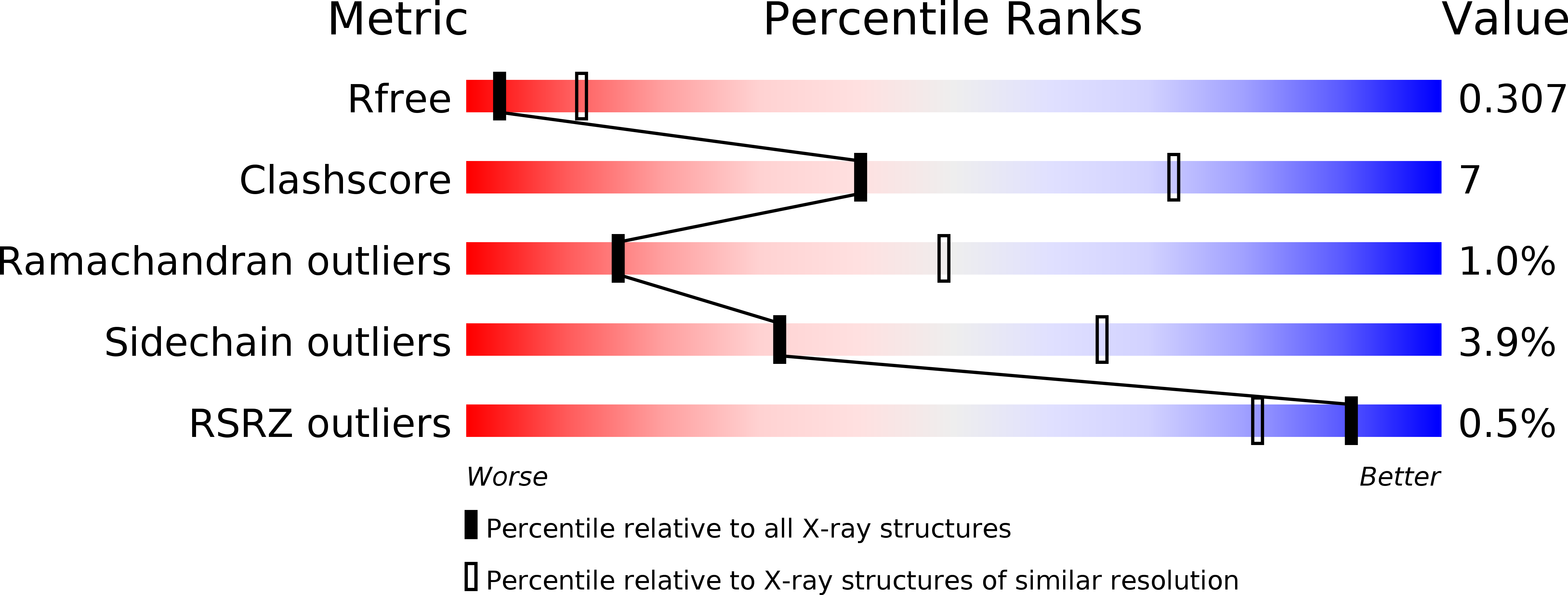
R-Value Free:
0.30
R-Value Work:
0.26
R-Value Observed:
0.26
Space Group:
C 1 2 1