6C0T image
Deposition Date 2018-01-02
Release Date 2018-05-30
Last Version Date 2024-10-09
Entry Detail
PDB ID:
6C0T
Title:
Crystal structure of cGMP-dependent protein kinase Ialpha (PKG Ialpha) catalytic domain bound with N46
Biological Source:
Source Organism:
Homo sapiens (Taxon ID: 9606)
Host Organism:
Method Details:
Experimental Method:
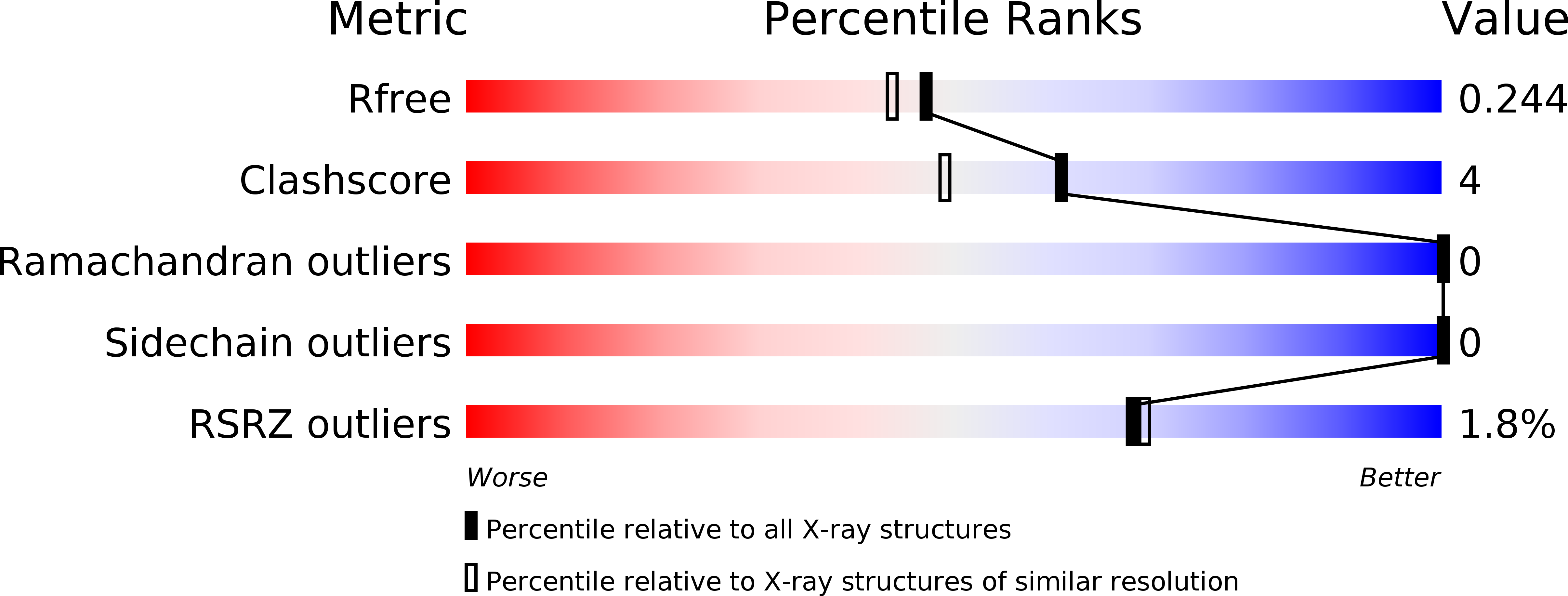
Resolution:
1.98 Å
R-Value Free:
0.24
R-Value Work:
0.20
R-Value Observed:
0.21
Space Group:
P 42
Macromolecular Entities
Polymer Type:polypeptide(L)
Molecule:cGMP-dependent protein kinase 1
Gene (Uniprot):PRKG1
Chain IDs:A
Chain Length:347
Number of Molecules:1
Biological Source:Homo sapiens
Modified Residue
Compound ID Chain ID Parent Comp ID Details 2D Image
TPO A THR modified residue
Primary Citation
Structural basis for selective inhibition of human PKG I alpha by the balanol-like compound N46.
J. Biol. Chem. 293 10985 10992 (2018)
PMID: 29769318 DOI: 10.1074/jbc.RA118.002427

Abstact

Activation of protein kinase G (PKG) Iα in nociceptive neurons induces long-term hyperexcitability that causes chronic pain. Recently, a derivative of the fungal metabolite balanol, N46, has been reported to inhibit PKG Iα with high potency and selectivity and attenuate thermal hyperalgesia and osteoarthritic pain. Here we determined co-crystal structures of the PKG Iα C-domain and cAMP-dependent protein kinase (PKA) Cα, each bound with N46, at 1.98 Å and 2.65 Å, respectively. N46 binds the active site with its external phenyl ring, specifically interacting with the glycine-rich loop and the αC helix. Phe-371 at the PKG Iα glycine-rich loop is oriented parallel to the phenyl ring of N46, forming a strong π-stacking interaction, whereas the analogous Phe-54 in PKA Cα rotates 30° and forms a weaker interaction. Structural comparison revealed that steric hindrance between the preceding Ser-53 and the propoxy group of the phenyl ring may explain the weaker interaction with PKA Cα. The analogous Gly-370 in PKG Iα, however, causes little steric hindrance with Phe-371. Moreover, Ile-406 on the αC helix forms a hydrophobic interaction with N46 whereas its counterpart in PKA, Thr-88, does not. Substituting these residues in PKG Iα with those in PKA Cα increases the IC50 values for N46, whereas replacing these residues in PKA Cα with those in PKG Iα reduces the IC50, consistent with our structural findings. In conclusion, our results explain the structural basis for N46-mediated selective inhibition of human PKG Iα and provide a starting point for structure-guided design of selective PKG Iα inhibitors.

Legend

Protein

Chemical

Disease

Primary Citation of related structures