
Deposition Date
2017-12-15
Release Date
2018-01-24
Last Version Date
2025-05-28
Entry Detail
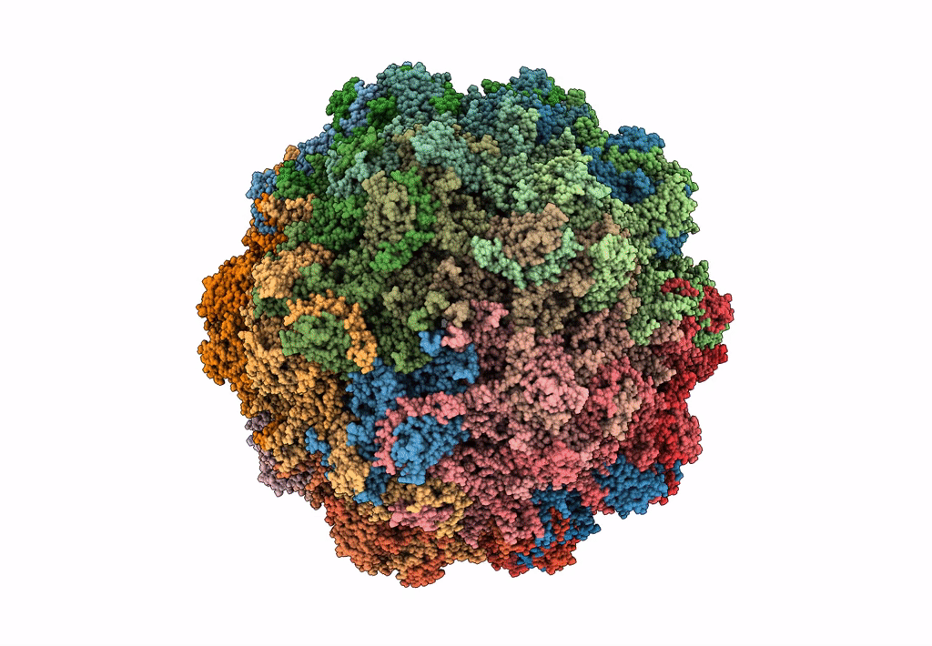
Biological Source:
Source Organism(s):
Bufavirus-1 (Taxon ID: 1209382)
Expression System(s):
Method Details:
Experimental Method:
Resolution:
2.84 Å
Aggregation State:
PARTICLE
Reconstruction Method:
SINGLE PARTICLE
