6BWK image
Deposition Date 2017-12-15
Release Date 2018-06-27
Last Version Date 2023-10-04
Entry Detail
PDB ID:
6BWK
Keywords:
Title:
Crystal structure of the human MLKL pseudokinase domain T357E/S358E mutant
Biological Source:
Source Organism:
Homo sapiens (Taxon ID: 9606)
Host Organism:
Method Details:
Experimental Method:
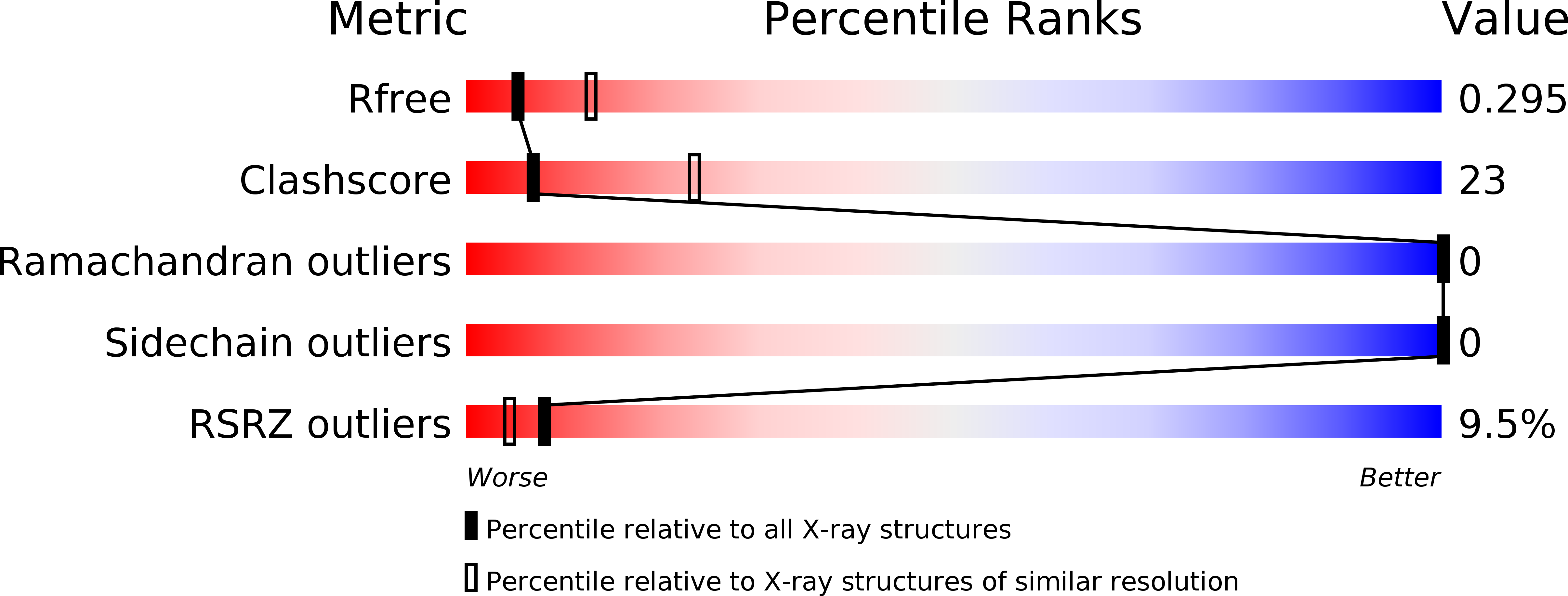
Resolution:
2.79 Å
R-Value Free:
0.29
R-Value Work:
0.25
R-Value Observed:
0.25
Space Group:
C 2 2 21
Macromolecular Entities
Structures with similar UniProt ID
Protein Blast
Polymer Type:polypeptide(L)
Molecule:Mixed lineage kinase domain-like protein
Gene (Uniprot):MLKL
Mutagens:T357E S358E
Chain IDs:A
Chain Length:287
Number of Molecules:1
Biological Source:Homo sapiens
Ligand Molecules
Primary Citation
Conformational switching of the pseudokinase domain promotes human MLKL tetramerization and cell death by necroptosis.
Nat Commun 9 2422 2422 (2018)
PMID: 29930286 DOI: 10.1038/s41467-018-04714-7

Abstact

Necroptotic cell death is mediated by the most terminal known effector of the pathway, MLKL. Precisely how phosphorylation of the MLKL pseudokinase domain activation loop by the upstream kinase, RIPK3, induces unmasking of the N-terminal executioner four-helix bundle (4HB) domain of MLKL, higher-order assemblies, and permeabilization of plasma membranes remains poorly understood. Here, we reveal the existence of a basal monomeric MLKL conformer present in human cells prior to exposure to a necroptotic stimulus. Following activation, toggling within the MLKL pseudokinase domain promotes 4HB domain disengagement from the pseudokinase domain αC helix and pseudocatalytic loop, to enable formation of a necroptosis-inducing tetramer. In contrast to mouse MLKL, substitution of RIPK3 substrate sites in the human MLKL pseudokinase domain completely abrogated necroptotic signaling. Therefore, while the pseudokinase domains of mouse and human MLKL function as molecular switches to control MLKL activation, the underlying mechanism differs between species.

Legend

Protein

Chemical

Disease

Primary Citation of related structures