6BUX image
Deposition Date 2017-12-11
Release Date 2018-07-18
Last Version Date 2023-10-04
Entry Detail
PDB ID:
6BUX
Keywords:
Title:
CRYSTAL STRUCTURE OF APOBEC3G CATALYTIC DOMAIN COMPLEX WITH SUBSTRATE SSDNA
Biological Source:
Source Organism:
Host Organism:
Method Details:
Experimental Method:
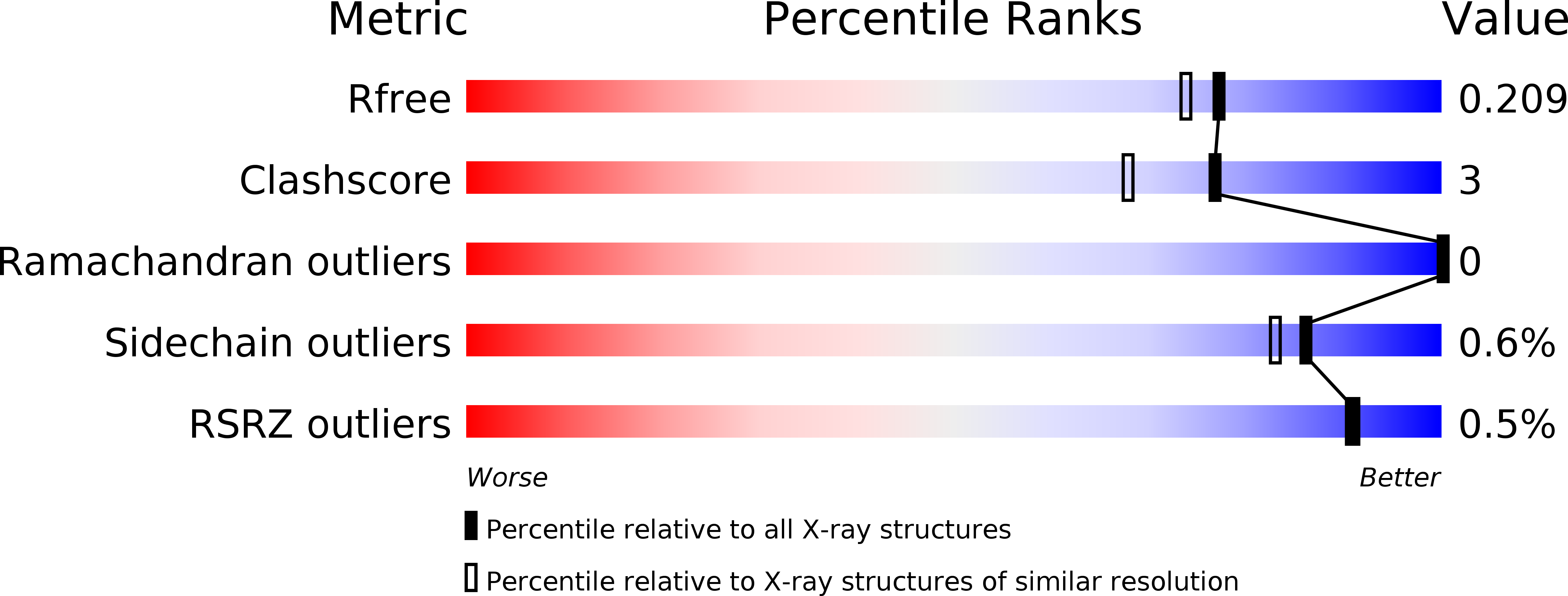
Resolution:
1.86 Å
R-Value Free:
0.21
R-Value Work:
0.17
R-Value Observed:
0.18
Space Group:
P 1 21 1
Macromolecular Entities
Polymer Type:polypeptide(L)
Molecule:Apolipoprotein B mRNA editing enzyme catalytic subunit 3G catalytic domain
Chain IDs:A
Chain Length:199
Number of Molecules:1
Biological Source:Homo sapiens
Polymer Type:polydeoxyribonucleotide
Molecule:DNA (5'-D(*AP*AP*TP*CP*CP*CP*AP*AP*A)-3')
Chain IDs:B
Chain Length:9
Number of Molecules:1
Biological Source:synthetic construct
Primary Citation
Crystal structure of the catalytic domain of HIV-1 restriction factor APOBEC3G in complex with ssDNA.
Nat Commun 9 2460 2460 (2018)
PMID: 29941968 DOI: 10.1038/s41467-018-04872-8

Abstact

The human APOBEC3G protein is a cytidine deaminase that generates cytidine to deoxy-uridine mutations in single-stranded DNA (ssDNA), and capable of restricting replication of HIV-1 by generating mutations in viral genome. The mechanism by which APOBEC3G specifically deaminates 5'-CC motifs has remained elusive since structural studies have been hampered due to apparently weak ssDNA binding of the catalytic domain of APOBEC3G. We overcame the problem by generating a highly active variant with higher ssDNA affinity. Here, we present the crystal structure of this variant complexed with a ssDNA substrate at 1.86 Å resolution. This structure reveals atomic-level interactions by which APOBEC3G recognizes a functionally-relevant 5'-TCCCA sequence. This complex also reveals a key role of W211 in substrate recognition, implicating a similar recognition in activation-induced cytidine deaminase (AID) with a conserved tryptophan.

Legend

Protein

Chemical

Disease

Primary Citation of related structures