6BTL image
Entry Detail
PDB ID:
6BTL
Title:
Crystal structure of Trypanothione Reductase from Trypanosoma brucei in complex with inhibitor RD117 1-[2-(Piperazin-1-yl)ethyl]-5-{5-[1-(pyrrolidin-1-yl)cyclohexyl]-1,3-thiazol-2-yl}-1H-indole
Biological Source:
Host Organism:
PDB Version:
Deposition Date:
2017-12-06
Release Date:
2018-06-06
Method Details:
Experimental Method:
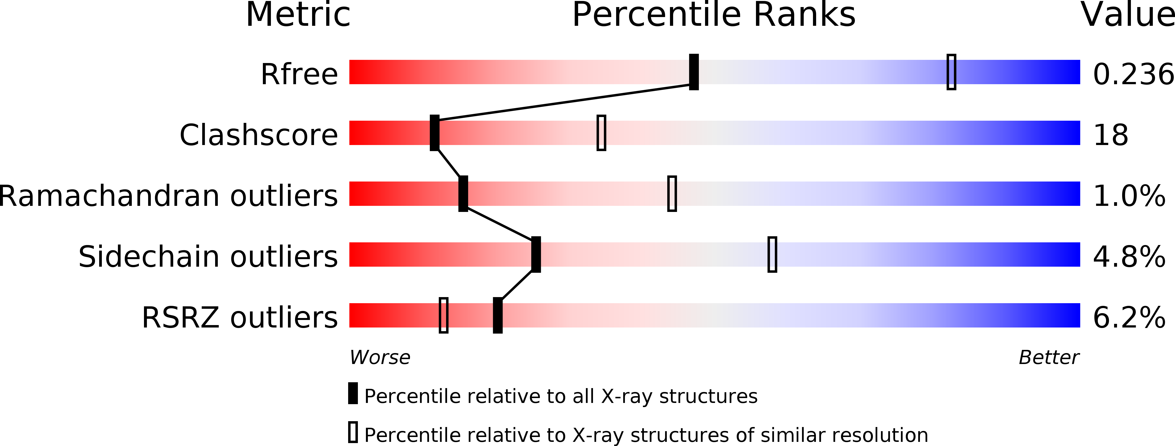
Resolution:
2.80 Å
R-Value Free:
0.23
R-Value Work:
0.19
R-Value Observed:
0.19
Space Group:
P 43 21 2
Macromolecular Entities
Polymer Type:polypeptide(L)
Description:Trypanothione reductase
Chain IDs:A, B
Chain Length:495
Number of Molecules:2
Biological Source:Trypanosoma brucei brucei TREU927
Primary Citation
Biological Evaluation and X-ray Co-crystal Structures of Cyclohexylpyrrolidine Ligands for Trypanothione Reductase, an Enzyme from the Redox Metabolism of Trypanosoma.
ChemMedChem 13 957 967 (2018)
PMID: 29624890 DOI: 10.1002/cmdc.201800067

Abstact

The tropical diseases human African trypanosomiasis, Chagas disease, and the various forms of leishmaniasis are caused by parasites of the family of trypanosomatids. These protozoa possess a unique redox metabolism based on trypanothione and trypanothione reductase (TR), making TR a promising drug target. We report the optimization of properties and potency of cyclohexylpyrrolidine inhibitors of TR by structure-based design. The best inhibitors were freely soluble and showed competitive inhibition constants (Ki) against Trypanosoma (T.) brucei TR and T. cruzi TR and in vitro activities (half-maximal inhibitory concentration, IC50) against these parasites in the low micromolar range, with high selectivity against human glutathione reductase. X-ray co-crystal structures confirmed the binding of the ligands to the hydrophobic wall of the "mepacrine binding site" with the new, solubility-providing vectors oriented toward the surface of the large active site.

Legend

Protein

Chemical

Disease

Primary Citation of related structures