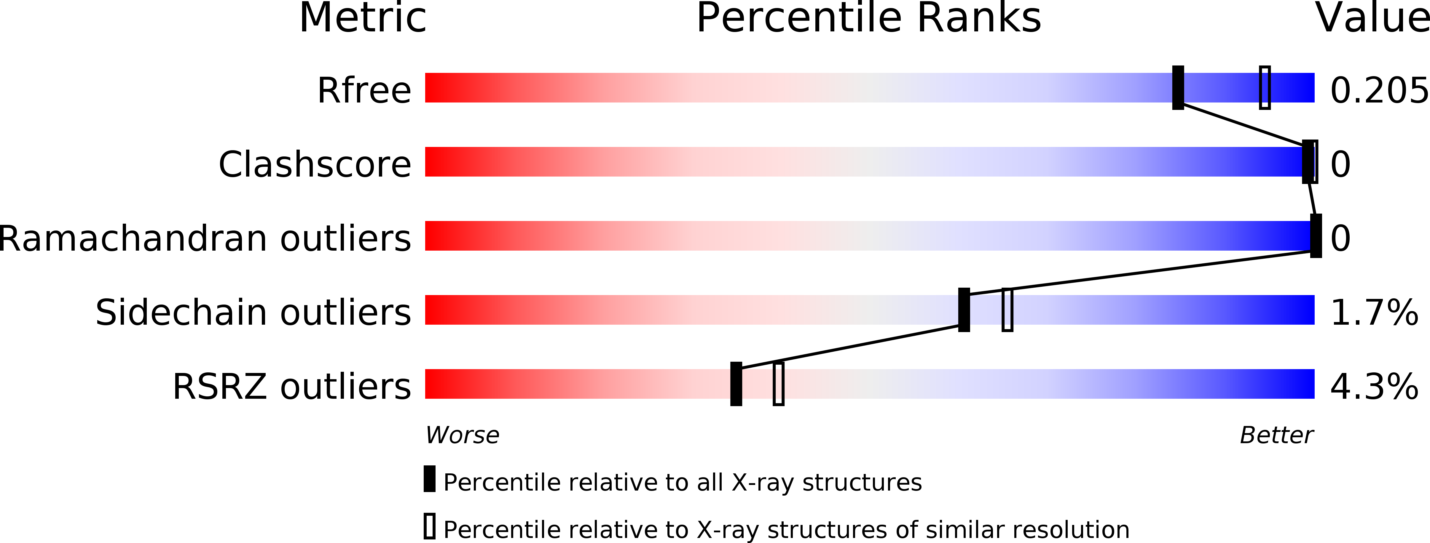
6BQC image
Entry Detail
PDB ID:
6BQC
Keywords:
Title:
Cyclopropane fatty acid synthase from E. coli
Biological Source:
Source Organism:
Host Organism:
PDB Version:
Deposition Date:
2017-11-27
Release Date:
2018-07-04
Method Details:
Experimental Method:
Resolution:
2.07 Å
R-Value Free:
0.20
R-Value Work:
0.17
R-Value Observed:
0.17
Space Group:
C 2 2 21
Macromolecular Entities
Polymer Type:polypeptide(L)
Description:Cyclopropane-fatty-acyl-phospholipid synthase
Chain IDs:A
Chain Length:383
Number of Molecules:1
Biological Source:Escherichia coli
Primary Citation
Structural and Functional Analysis of E. coli Cyclopropane Fatty Acid Synthase.
Structure 26 1251 ? (2018)
PMID: 30057024 DOI: 10.1016/j.str.2018.06.008

Abstact

Cell membranes must adapt to different environments. In Gram-negative bacteria, the inner membrane can be remodeled directly by modification of lipids embedded in the bilayer. For example, when Escherichia coli enters stationary phase, cyclopropane fatty acid (CFA) synthase converts most double bonds in unsaturated inner-membrane lipids into cyclopropyl groups. Here we report the crystal structure of E. coli CFA synthase. The enzyme is a dimer in the crystal and in solution, with each subunit containing a smaller N-domain that associates tightly with a larger catalytic C-domain, even following cleavage of the inter-domain linker or co-expression of each individual domain. Efficient catalysis requires dimerization and proper linkage of the two domains. These findings support an avidity-based model in which one subunit of the dimer stabilizes membrane binding, while the other subunit carries out catalysis.

Legend

Protein

Chemical

Disease

Primary Citation of related structures