6BOU image
Deposition Date 2017-11-20
Release Date 2018-08-15
Last Version Date 2023-10-04
Entry Detail
PDB ID:
6BOU
Title:
Human APE1 substrate complex with an T/C mismatch adjacent the THF
Biological Source:
Source Organism:
Host Organism:
Method Details:
Experimental Method:
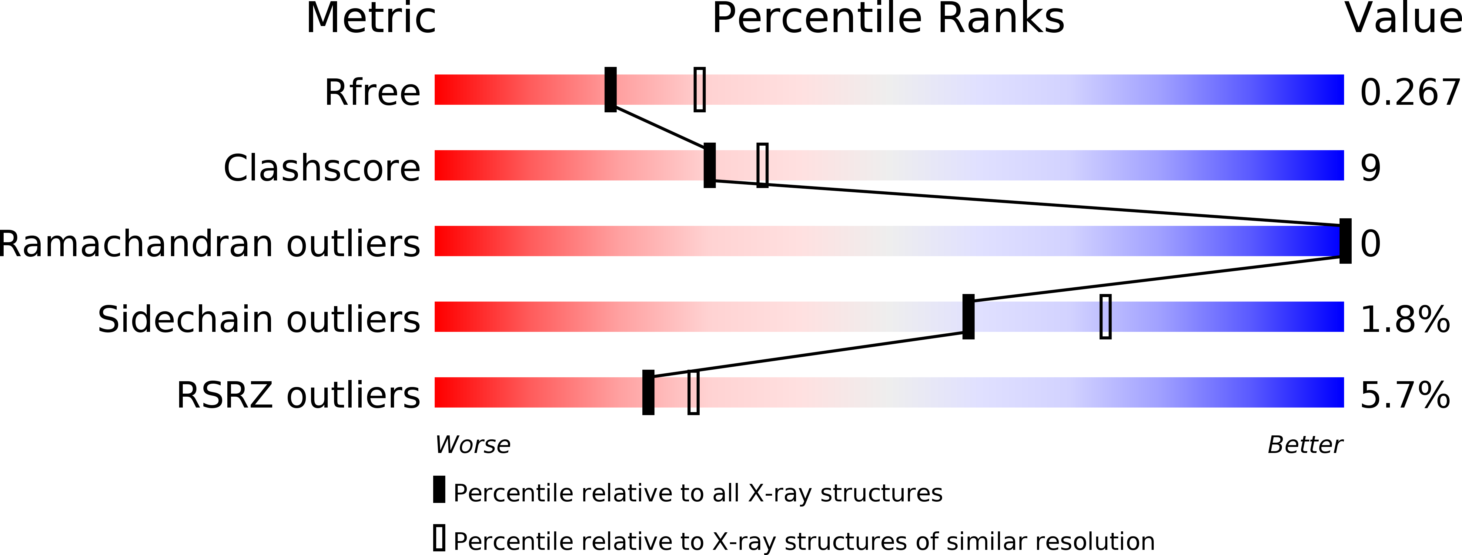
Resolution:
2.54 Å
R-Value Free:
0.26
R-Value Work:
0.20
R-Value Observed:
0.21
Space Group:
P 1
Macromolecular Entities
Polymer Type:polypeptide(L)
Molecule:DNA-(apurinic or apyrimidinic site) lyase
Gene (Uniprot):APEX1
Mutations:C138A
Chain IDs:A, B
Chain Length:318
Number of Molecules:2
Biological Source:Homo sapiens
Polymer Type:polydeoxyribonucleotide
Molecule:21-mer DNA
Chain IDs:C (auth: P)
Chain Length:21
Number of Molecules:1
Biological Source:synthetic construct
Polymer Type:polydeoxyribonucleotide
Molecule:21-mer DNA
Chain IDs:D (auth: V)
Chain Length:21
Number of Molecules:1
Biological Source:synthetic construct
Primary Citation
Apurinic/apyrimidinic (AP) endonuclease 1 processing of AP sites with 5' mismatches.
Acta Crystallogr D Struct Biol 74 760 768 (2018)
PMID: 30082511 DOI: 10.1107/S2059798318003340

Abstact

Despite the DNA duplex being central to biological functions, many intricacies of this molecule, including the dynamic nature of mismatched base pairing, are still unknown. The unique conformations adopted by DNA mismatches can provide insight into the forces at play between nucleotides. Moreover, DNA-binding proteins apply their own individualized steric and electrochemical influences on the nucleotides that they interact with, further altering base-pairing conformations. Here, seven X-ray crystallographic structures of the human nuclease apurinic/apyrimidinic (AP) endonuclease 1 (APE1) in complex with its substrate target flanked by a 5' mismatch are reported. The structures reveal how APE1 influences the conformations of a variety of different mismatched base pairs. Purine-purine mismatches containing a guanine are stabilized by a rotation of the guanine residue about the N-glycosidic bond to utilize the Hoogsteen edge for hydrogen bonding. Interestingly, no rotation of adenine, the other purine, is observed. Mismatches involving both purine and pyrimidine bases adopt wobble conformations to accommodate the mismatch. Pyrimidine-pyrimidine mismatches also wobble; however, the smaller profile of a pyrimidine base results in a gap between the Watson-Crick faces that is reduced by a C1'-C1' compression. These results advance our understanding of mismatched base pairing and the influence of a bound protein.

Legend

Protein

Chemical

Disease

Primary Citation of related structures