6BNN image
Entry Detail
PDB ID:
6BNN
Keywords:
Title:
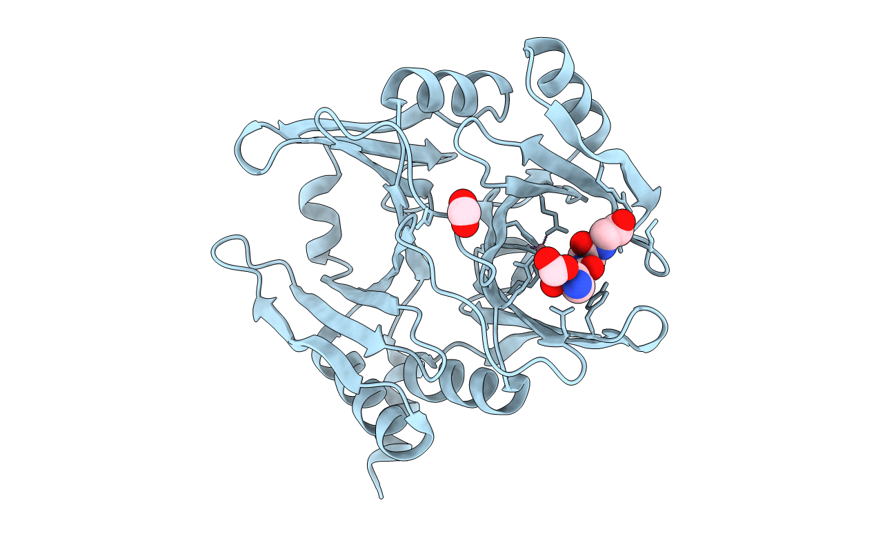
Crystal structure of V278E-glyoxalase I mutant from Zea mays in space group P4(1)2(1)2
Biological Source:
Source Organism:
Host Organism:
PDB Version:
Deposition Date:
2017-11-17
Release Date:
2018-11-21
Method Details:
Experimental Method:
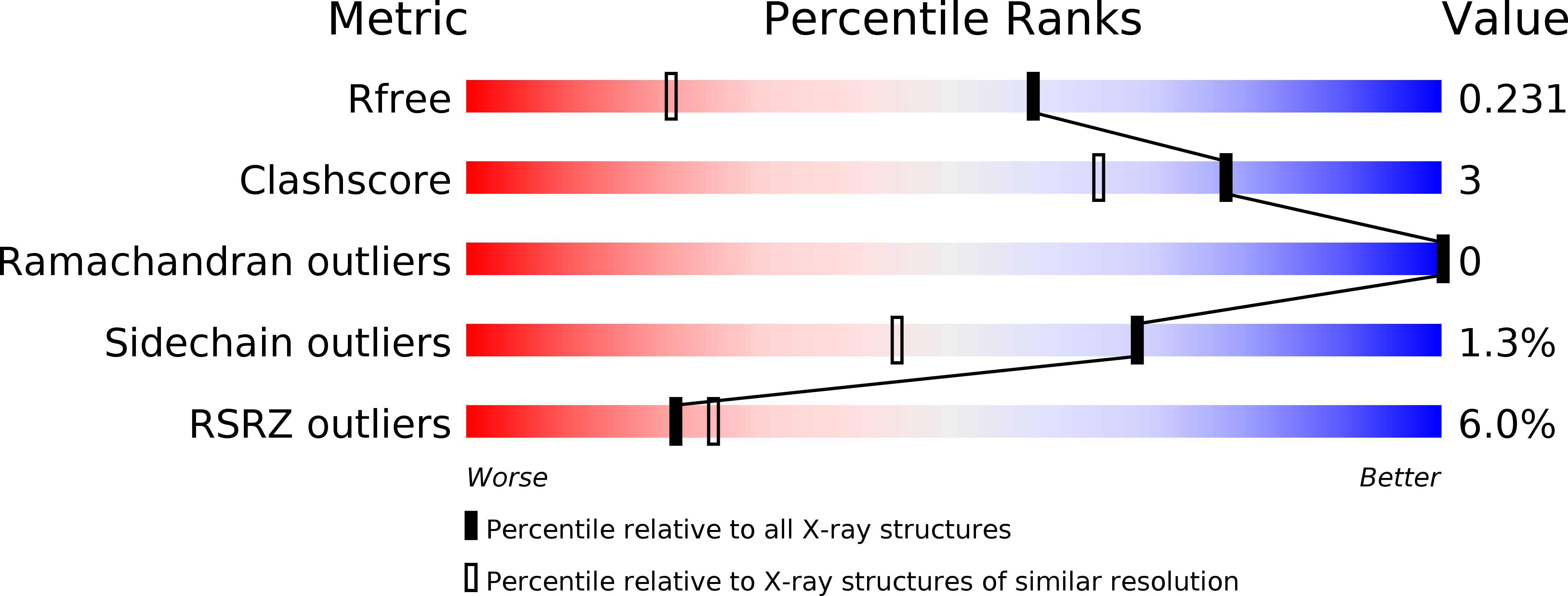
Resolution:
1.55 Å
R-Value Free:
0.23
R-Value Work:
0.18
R-Value Observed:
0.19
Space Group:
P 41 21 2
Macromolecular Entities
Polymer Type:polypeptide(L)
Description:Lactoylglutathione lyase
Mutations:V278E
Chain IDs:A
Chain Length:296
Number of Molecules:1
Biological Source:Zea mays
Primary Citation
Deciphering the number and location of active sites in the monomeric glyoxalase I of Zea mays.
Febs J. 286 3255 3271 (2019)
PMID: 30993890 DOI: 10.1111/febs.14855

Abstact

Detoxification of methylglyoxal, a toxic by-product of central sugar metabolism, is a major issue for all forms of life. The glyoxalase pathway evolved to effectively convert methylglyoxal into d-lactate via a glutathione hemithioacetal intermediate. Recently, we have shown that the monomeric glyoxalase I from maize exhibits a symmetric fold with two cavities, potentially harboring two active sites, in analogy with homodimeric enzyme surrogates. Here we confirm that only one of the two cavities exhibits glyoxalase I activity and show that it adopts a tunnel-shaped structure upon substrate binding. Such conformational change gives rise to independent binding sites for glutathione and methylglyoxal in the same active site, with important implications for the molecular reaction mechanism, which has been a matter of debate for several decades. DATABASE: Structural data are available in The Protein Data Bank database under the accession numbers 6BNN, 6BNX, and 6BNZ.

Legend

Protein

Chemical

Disease

Primary Citation of related structures