Deposition Date
2017-11-14
Release Date
2017-12-06
Last Version Date
2025-05-28
Entry Detail
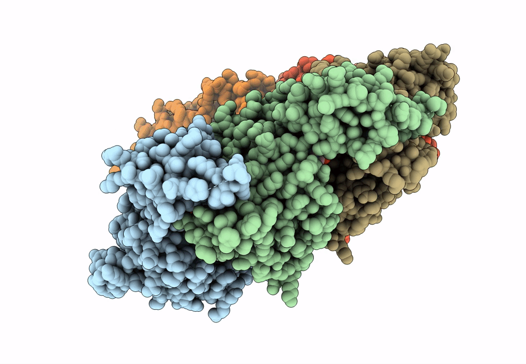
PDB ID:
6BMF
Keywords:
Title:
Vps4p-Vta1p complex with peptide binding to the central pore of Vps4p
Biological Source:
Source Organism(s):
Saccharomyces cerevisiae (Taxon ID: 4932)
Expression System(s):
Method Details:
Experimental Method:
Resolution:
3.20 Å
Aggregation State:
PARTICLE
Reconstruction Method:
SINGLE PARTICLE