6BKA image
Deposition Date 2017-11-08
Release Date 2018-02-14
Last Version Date 2024-11-06
Entry Detail
PDB ID:
6BKA
Keywords:
Title:
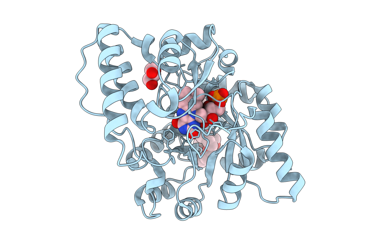
Crystal Structure of Nitronate Monooxygenase from Cyberlindnera saturnus
Biological Source:
Source Organism:
Host Organism:
Method Details:
Experimental Method:
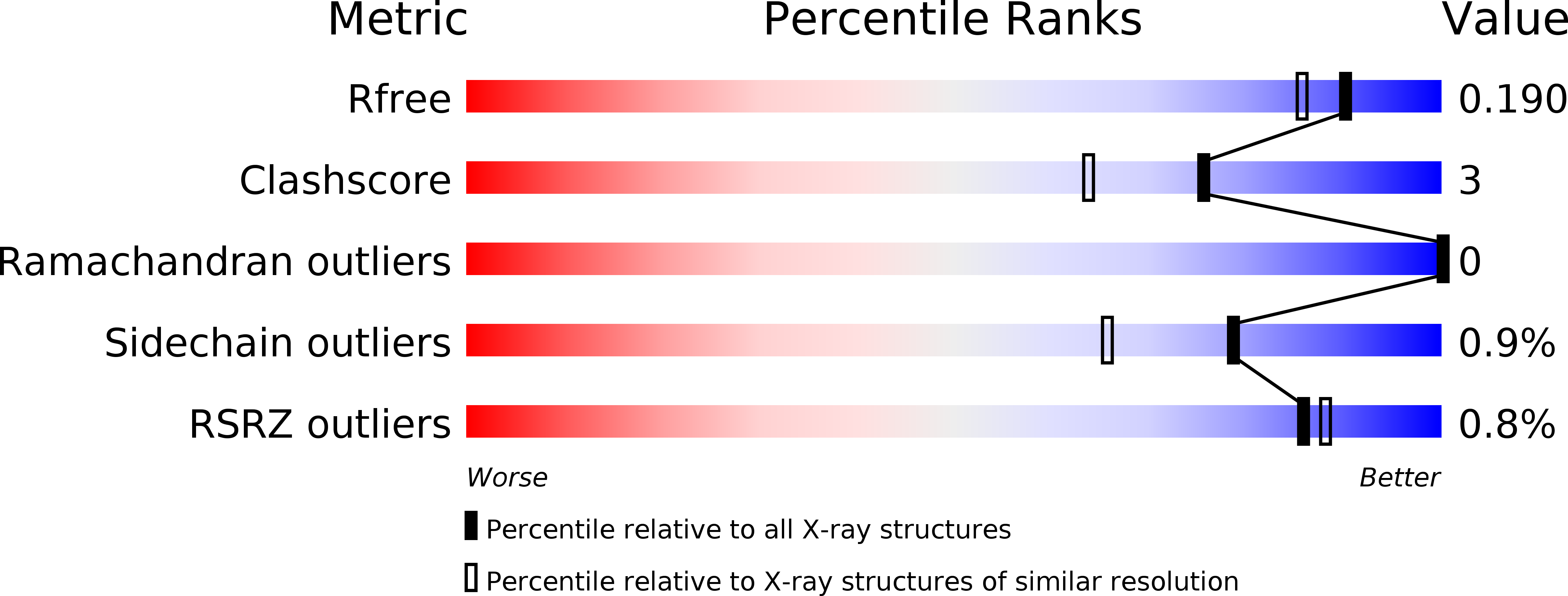
Resolution:
1.65 Å
R-Value Free:
0.17
R-Value Work:
0.13
R-Value Observed:
0.14
Space Group:
P 1 21 1
Macromolecular Entities
Polymer Type:polypeptide(L)
Molecule:Nitronate monooxygenase
Chain IDs:A
Chain Length:374
Number of Molecules:1
Biological Source:Cyberlindnera mrakii
Modified Residue
Compound ID Chain ID Parent Comp ID Details 2D Image
CSD A CYS modified residue
Primary Citation
Crystal structure of yeast nitronate monooxygenase from Cyberlindnera saturnus.
Proteins 86 599 605 (2018)
PMID: 29383742 DOI: 10.1002/prot.25470

Abstact

Nitronate monooxygenase (NMO) is an FMN-dependent enzyme that oxidizes the neurotoxin propionate 3-nitronate (P3N) and represents the best-known system for P3N detoxification in different organisms. The crystal structure of the first eukaryotic Class I NMO from Cyberlindnera saturnus (CsNMO) has been solved at 1.65 Å resolution and refined to an R-factor of 14.0%. The three-dimensional structures of yeast CsNMO and bacterial PaNMO are highly conserved with the exception of three additional loops on the surface in the CsNMO enzyme and differences in four active sites residues. A PEG molecule was identified in the structure and formed extensive interactions with CsNMO, suggesting a specific binding site; however, 8% PEG showed no significant effect on the enzyme activity. This new crystal structure of a eukaryotic NMO provides insight into the function of this class of enzymes.

Legend

Protein

Chemical

Disease

Primary Citation of related structures