6BGD image
Deposition Date 2017-10-27
Release Date 2018-01-31
Last Version Date 2023-10-25
Entry Detail
PDB ID:
6BGD
Title:
The crystal structure of the W145A variant of TpMglB-2 (Tp0684) with bound ligand
Biological Source:
Source Organism:
Host Organism:
Method Details:
Experimental Method:
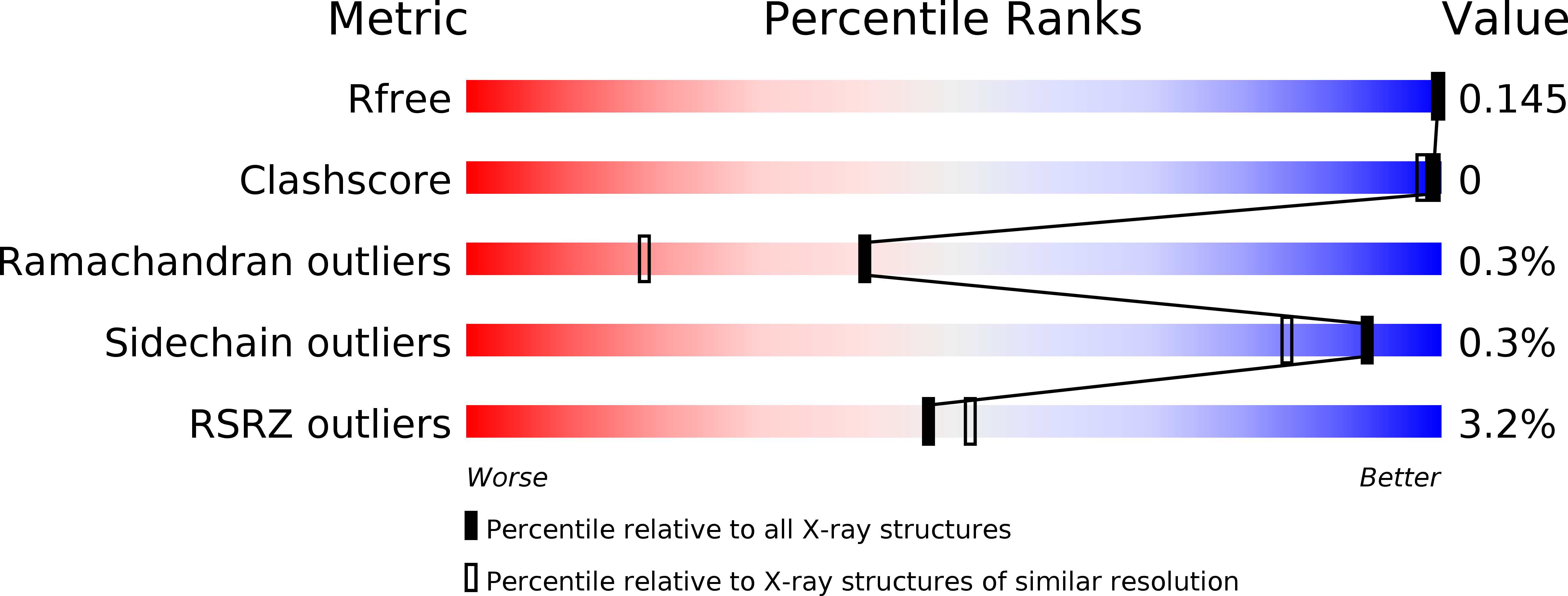
Resolution:
1.47 Å
R-Value Free:
0.14
R-Value Work:
0.11
R-Value Observed:
0.11
Space Group:
C 2 2 21
Macromolecular Entities
Structures with similar UniProt ID
Protein Blast
Polymer Type:polypeptide(L)
Molecule:Glucose/galactose-binding lipoprotein
Gene (Uniprot):mglB
Chain IDs:A
Chain Length:378
Number of Molecules:1
Biological Source:Treponema pallidum (strain Nichols)
Primary Citation
Crystal structures of MglB-2 (TP0684), a topologically variant d-glucose-binding protein from Treponema pallidum, reveal a ligand-induced conformational change.
Protein Sci. 27 880 885 (2018)
PMID: 29318719 DOI: 10.1002/pro.3373

Abstact

Previously, we determined the crystal structure of apo-TpMglB-2, a d-glucose-binding component of a putative ABC transporter from the syphilis spirochete Treponema pallidum. The protein had an unusual topology for this class of proteins, raising the question of whether the d-glucose-binding mode would be different in TpMglB-2. Here, we present the crystal structures of a variant of TpMglB-2 with and without d-glucose bound. The structures demonstrate that, despite its aberrant topology, the protein undergoes conformational changes and binds d-glucose similarly to other Mgl-type proteins, likely facilitating d-glucose uptake in T. pallidum.

Legend

Protein

Chemical

Disease

Primary Citation of related structures