6BDE image
Deposition Date 2017-10-22
Release Date 2018-05-30
Last Version Date 2023-10-04
Entry Detail
PDB ID:
6BDE
Title:
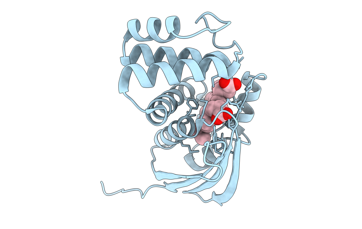
Crystal structure of Fe(II) unliganded H-NOX protein mutant A71G from K. algicida
Biological Source:
Source Organism:
Host Organism:
Method Details:
Experimental Method:
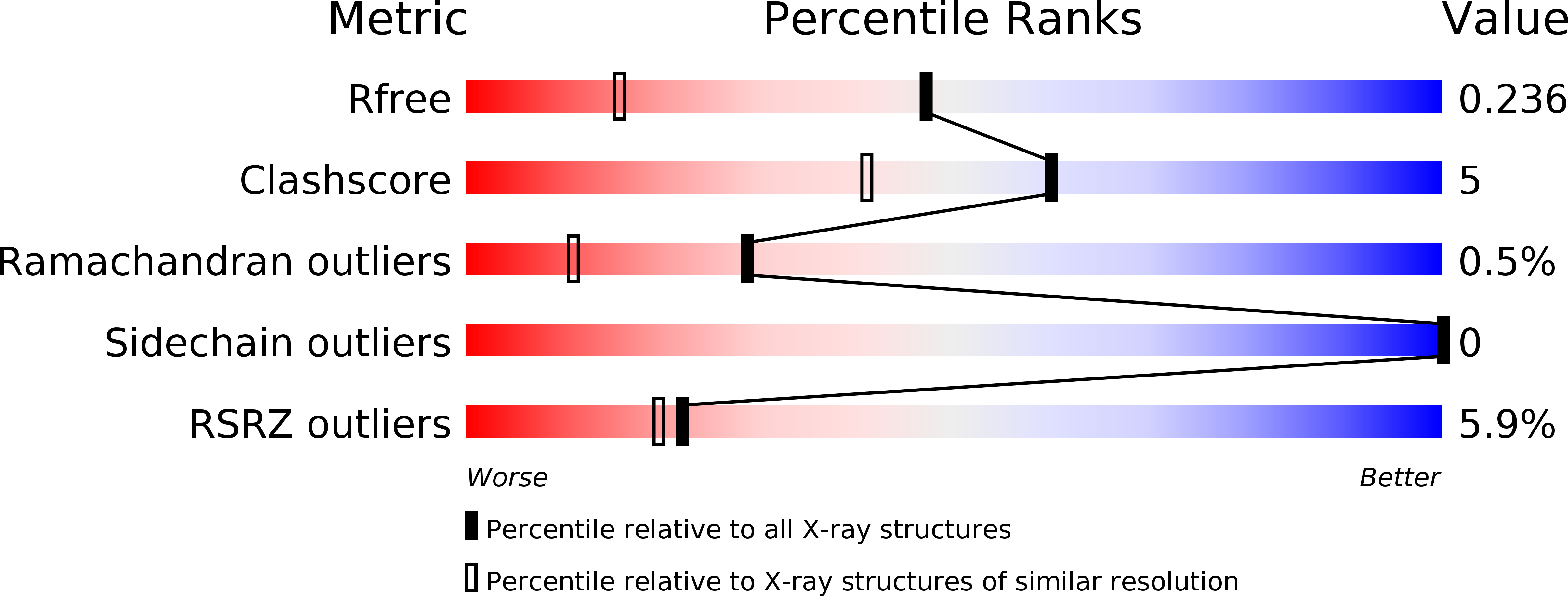
Resolution:
1.64 Å
R-Value Free:
0.23
R-Value Work:
0.21
R-Value Observed:
0.21
Space Group:
P 21 21 21
Macromolecular Entities
Structures with similar UniProt ID
Protein Blast
Polymer Type:polypeptide(L)
Molecule:2,4-dihydroxyhept-2-ene-1,7-dioic acid aldolase
Gene (Uniprot):KAOT1_17313
Mutagens:A71G
Chain IDs:A
Chain Length:185
Number of Molecules:1
Biological Source:Kordia algicida OT-1
Ligand Molecules
Primary Citation
Native Alanine Substitution in the Glycine Hinge Modulates Conformational Flexibility of Heme Nitric Oxide/Oxygen (H-NOX) Sensing Proteins.
ACS Chem. Biol. 13 1631 1639 (2018)
PMID: 29757599 DOI: 10.1021/acschembio.8b00248

Abstact

Heme nitric oxide/oxygen sensing (H-NOX) domains are direct NO sensors that regulate a variety of biological functions in both bacteria and eukaryotes. Previous work on H-NOX proteins has shown that upon NO binding, a conformational change occurs along two glycine residues on adjacent helices (termed the glycine hinge). Despite the apparent importance of the glycine hinge, it is not fully conserved in all H-NOX domains. Several H-NOX sensors from the family Flavobacteriaceae contain a native alanine substitution in one of the hinge residues. In this work, the effect of the increased steric bulk within the Ala-Gly hinge on H-NOX function was investigated. The hinge in Kordia algicida OT-1 (Ka H-NOX) is composed of A71 and G145. Ligand-binding properties and signaling function for this H-NOX were characterized. The variant A71G was designed to convert the hinge region of Ka H-NOX to the typical Gly-Gly motif. In activity assays with its cognate histidine kinase (HnoK), the wild type displayed increased signal specificity compared to A71G. Increasing titrations of unliganded A71G gradually inhibits HnoK autophosphorylation, while increasing titrations of unliganded wild type H-NOX does not inhibit HnoK. Crystal structures of both wild type and A71G Ka H-NOX were solved to 1.9 and 1.6 Å, respectively. Regions of H-NOX domains previously identified as involved in protein-protein interactions with HnoK display significantly higher b-factors in A71G compared to wild-type H-NOX. Both biochemical and structural data indicate that the hinge region controls overall conformational flexibility of the H-NOX, affecting NO complex formation and regulation of its HnoK.

Legend

Protein

Chemical

Disease

Primary Citation of related structures
Feedback Form
Name
Email
Institute
Feedback