6BC9 image
Entry Detail
PDB ID:
6BC9
Title:
Joint X-ray/neutron structure of human carbonic anhydrase II in complex with dorzolamide
Biological Source:
Source Organism:
Host Organism:
PDB Version:
Deposition Date:
2017-10-20
Release Date:
2018-02-28
Method Details:
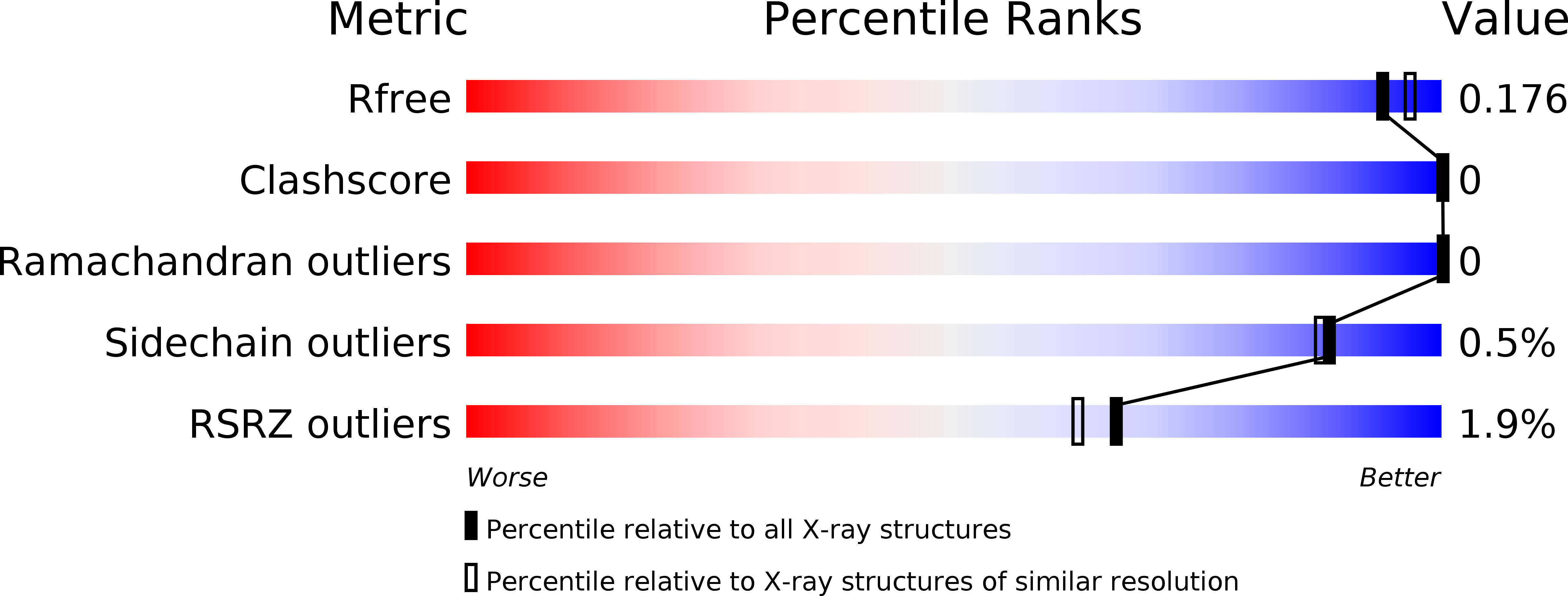
R-Value Free:
['0.19
R-Value Work:
['0.17
R-Value Observed:
['?', '?'].00
Space Group:
P 1 21 1
Macromolecular Entities
Polymer Type:polypeptide(L)
Description:Carbonic anhydrase 2
Chain IDs:A
Chain Length:260
Number of Molecules:1
Biological Source:Homo sapiens
Primary Citation
"To Be or Not to Be" Protonated: Atomic Details of Human Carbonic Anhydrase-Clinical Drug Complexes by Neutron Crystallography and Simulation.
Structure 26 383 390.e3 (2018)
PMID: 29429876 DOI: 10.1016/j.str.2018.01.006

Abstact

Human carbonic anhydrases (hCAs) play various roles in cells, and have been drug targets for decades. Sequence similarities of hCA isoforms necessitate designing specific inhibitors, which requires detailed structural information for hCA-inhibitor complexes. We present room temperature neutron structures of hCA II in complex with three clinical drugs that provide in-depth analysis of drug binding, including protonation states of the inhibitors, hydration water structure, and direct visualization of hydrogen-bonding networks in the enzyme's active site. All sulfonamide inhibitors studied bind to the Zn metal center in the deprotonated, anionic, form. Other chemical groups of the drugs can remain neutral or be protonated when bound to hCA II. MD simulations have shown that flexible functional groups of the inhibitors may alter their conformations at room temperature and occupy different sub-sites. This study offers insights into the design of specific drugs to target cancer-related hCA isoform IX.

Legend

Protein

Chemical

Disease

Primary Citation of related structures