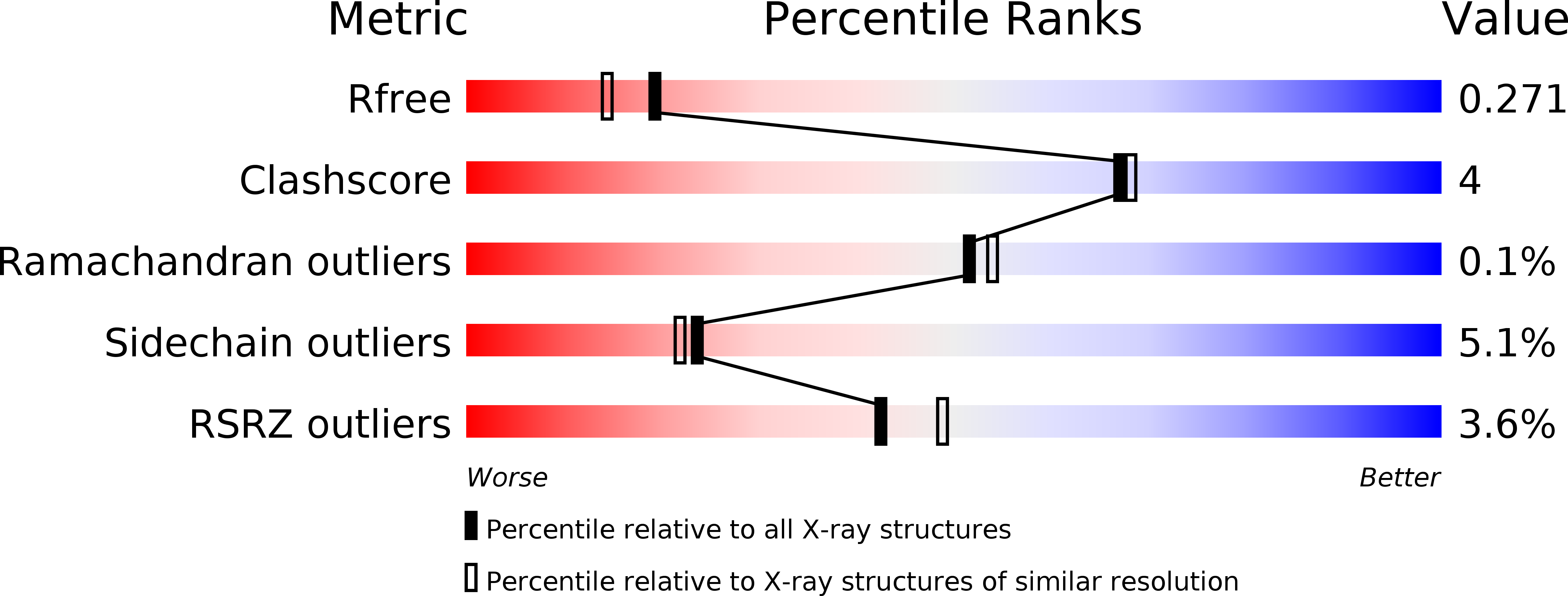
6BB4 image
Deposition Date 2017-10-16
Release Date 2018-05-02
Last Version Date 2024-10-30
Entry Detail
PDB ID:
6BB4
Keywords:
Title:
Fab/epitope complex of mouse monoclonal antibody C5.2 targeting a phospho-tau epitope.
Biological Source:
Source Organism:
Homo sapiens (Taxon ID: 9606)
Mus musculus (Taxon ID: 10090)
Method Details:
Experimental Method:
Resolution:
2.10 Å
R-Value Free:
0.26
R-Value Work:
0.21
Space Group:
C 1 2 1
Macromolecular Entities
Polymer Type:polypeptide(L)
Molecule:Mouse monoclonal antibody C5.2 Fab heavy chain
Chain IDs:B (auth: H), E (auth: I), H (auth: J)
Chain Length:220
Number of Molecules:3
Biological Source:Mus musculus
Polymer Type:polypeptide(L)
Molecule:Mouse monoclonal antibody C5.2 Fab light chain
Chain IDs:A (auth: L), D (auth: M), G (auth: N)
Chain Length:213
Number of Molecules:3
Biological Source:Mus musculus
Structures with similar UniProt ID
Protein Blast
Polymer Type:polypeptide(L)
Molecule:Microtubule-associated protein tau
Gene (Uniprot):MAPT
Chain IDs:C (auth: P), F (auth: Q), I (auth: R)
Chain Length:23
Number of Molecules:3
Biological Source:Homo sapiens
Modified Residue
Compound ID Chain ID Parent Comp ID Details 2D Image
SEP C SER modified residue
Ligand Molecules
Primary Citation
Tau Antibody Structure Reveals a Molecular Switch Defining a Pathological Conformation of the Tau Protein.
Sci Rep 8 6209 6209 (2018)
PMID: 29670132 DOI: 10.1038/s41598-018-24276-4

Abstact

Tau antibodies have shown therapeutic potential for Alzheimer's disease and several are in clinical trials. As a microtubule-associated protein, tau relies on dynamic phosphorylation for its normal functions. In tauopathies, it becomes hyperphosphorylated and aggregates into toxic assemblies, which collectively lead to neurodegeneration. Of the phospho-epitopes, the region around Ser396 has received particular attention because of its prominence and stability in tauopathies. Here we report the first structure of a monoclonal tau antibody in complex with the pathologically important phospho-Ser396 residue. Its binding region reveals tau residues Tyr394 to phospho-Ser396 stabilized in a β-strand conformation that is coordinated by a phospho-specific antigen binding site. These details highlight a molecular switch that defines this prominent conformation of tau and ways to target it. Overall, the structure of the antibody-antigen complex clarifies why certain phosphorylation sites in tau are more closely linked to neurodegeneration than others.

Legend

Protein

Chemical

Disease

Primary Citation of related structures