6BAJ image
Entry Detail
PDB ID:
6BAJ
EMDB ID:
Title:
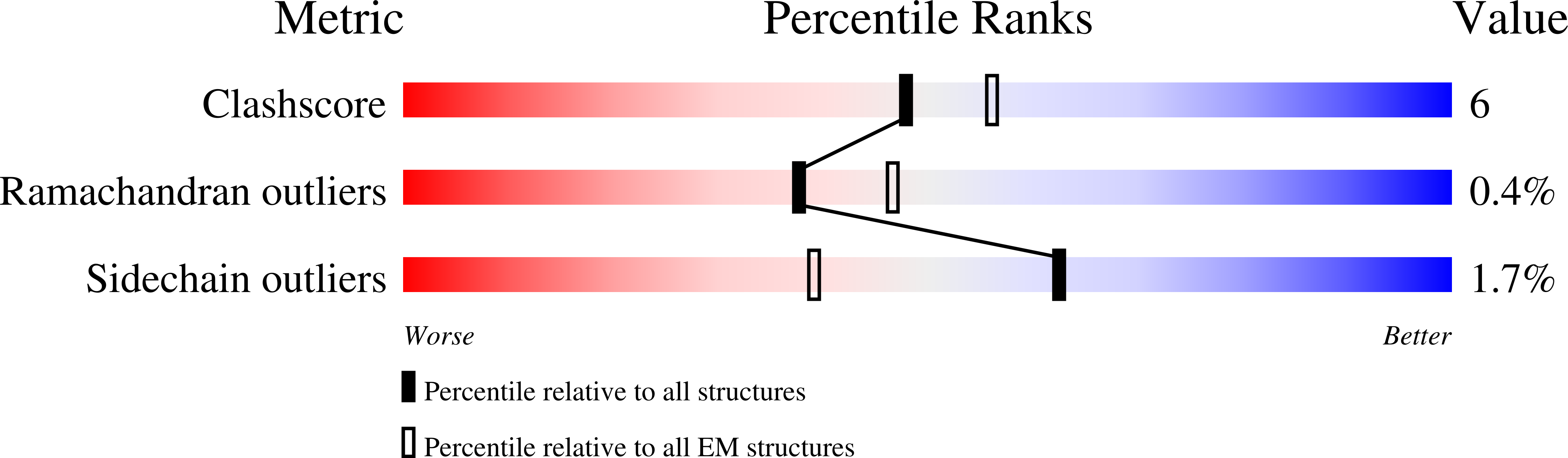
Cryo-EM structure of lipid bilayer in the native cell membrane nanoparticles of AcrB
Biological Source:
Host Organism:
PDB Version:
Deposition Date:
2017-10-13
Release Date:
2018-12-19
Method Details:
Experimental Method:
Resolution:
3.20 Å
Aggregation State:
PARTICLE
Reconstruction Method:
SINGLE PARTICLE
Macromolecular Entities
Polymer Type:polypeptide(L)
Description:Multidrug efflux pump subunit AcrB
Chain IDs:A (auth: B), B (auth: C), C (auth: A)
Chain Length:1057
Number of Molecules:3
Biological Source:Escherichia coli (strain K12)
Primary Citation
Structure and activity of lipid bilayer within a membrane-protein transporter.
Proc.Natl.Acad.Sci.USA 115 12985 12990 (2018)
PMID: 30509977 DOI: 10.1073/pnas.1812526115

Abstact

Membrane proteins function in native cell membranes, but extraction into isolated particles is needed for many biochemical and structural analyses. Commonly used detergent-extraction methods destroy naturally associated lipid bilayers. Here, we devised a detergent-free method for preparing cell-membrane nanoparticles to study the multidrug exporter AcrB, by cryo-EM at 3.2-Å resolution. We discovered a remarkably well-organized lipid-bilayer structure associated with transmembrane domains of the AcrB trimer. This bilayer patch comprises 24 lipid molecules; inner leaflet chains are packed in a hexagonal array, whereas the outer leaflet has highly irregular but ordered packing. Protein side chains interact with both leaflets and participate in the hexagonal pattern. We suggest that the lipid bilayer supports and harmonizes peristaltic motions through AcrB trimers. In AcrB D407A, a putative proton-relay mutant, lipid bilayer buttresses protein interactions lost in crystal structures after detergent-solubilization. Our detergent-free system preserves lipid-protein interactions for visualization and should be broadly applicable.

Legend

Protein

Chemical

Disease

Primary Citation of related structures