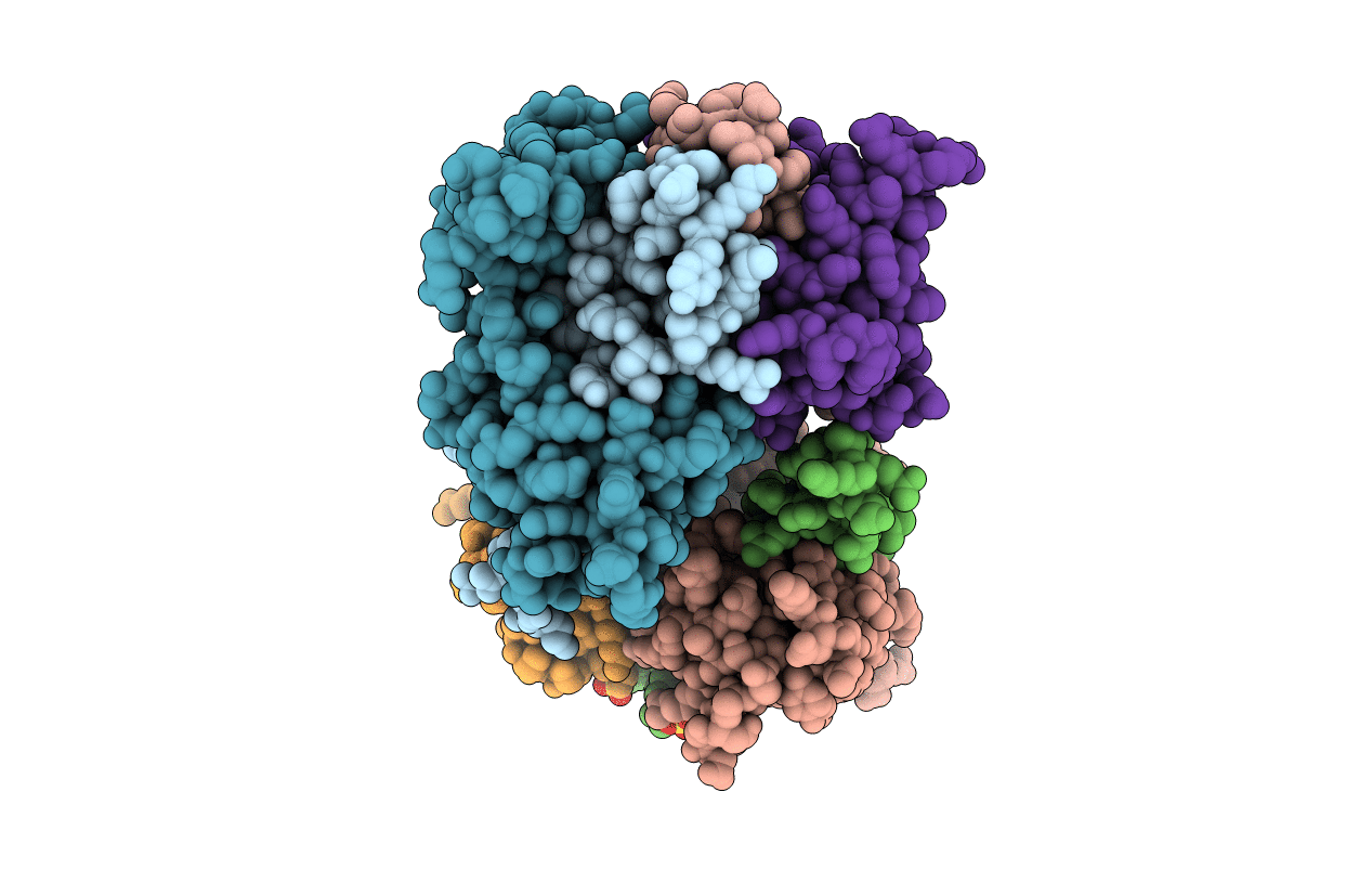
6B8N image
Entry Detail
PDB ID:
6B8N
Keywords:
Title:
Crystal Structure of the Ca2+/CaM:Kv7.4 (KCNQ4) AB Domain Complex, 10 uM CaCl2 soak
Biological Source:
Source Organism:
Host Organism:
PDB Version:
Deposition Date:
2017-10-09
Release Date:
2018-03-14
Method Details:
Experimental Method:
Resolution:
2.20 Å
R-Value Free:
0.26
R-Value Work:
0.22
R-Value Observed:
0.22
Space Group:
I 2 2 2
Macromolecular Entities
Polymer Type:polypeptide(L)
Description:Potassium voltage-gated channel subfamily KQT member 4
Chain IDs:A, C, E, G
Chain Length:82
Number of Molecules:4
Biological Source:Homo sapiens
Polymer Type:polypeptide(L)
Description:Calmodulin-1
Chain IDs:B, D, F, H
Chain Length:149
Number of Molecules:4
Biological Source:Homo sapiens
Primary Citation
A Calmodulin C-Lobe Ca
Neuron 97 836 852.e6 (2018)
PMID: 29429937 DOI: 10.1016/j.neuron.2018.01.035

Abstact

Kv7 (KCNQ) voltage-gated potassium channels control excitability in the brain, heart, and ear. Calmodulin (CaM) is crucial for Kv7 function, but how this calcium sensor affects activity has remained unclear. Here, we present X-ray crystallographic analysis of CaM:Kv7.4 and CaM:Kv7.5 AB domain complexes that reveal an Apo/CaM clamp conformation and calcium binding preferences. These structures, combined with small-angle X-ray scattering, biochemical, and functional studies, establish a regulatory mechanism for Kv7 CaM modulation based on a common architecture in which a CaM C-lobe calcium-dependent switch releases a shared Apo/CaM clamp conformation. This C-lobe switch inhibits voltage-dependent activation of Kv7.4 and Kv7.5 but facilitates Kv7.1, demonstrating that mechanism is shared by Kv7 isoforms despite the different directions of CaM modulation. Our findings provide a unified framework for understanding how CaM controls different Kv7 isoforms and highlight the role of membrane proximal domains for controlling voltage-gated channel function. VIDEO ABSTRACT.

Legend

Protein

Chemical

Disease

Primary Citation of related structures