6B7M image
Deposition Date 2017-10-04
Release Date 2018-04-18
Last Version Date 2024-03-13
Entry Detail
PDB ID:
6B7M
Keywords:
Title:
Crystal structure of Legionella effector sdeD (lpg2509) in complex with Ubiquitin
Biological Source:
Source Organism:
Host Organism:
Method Details:
Experimental Method:
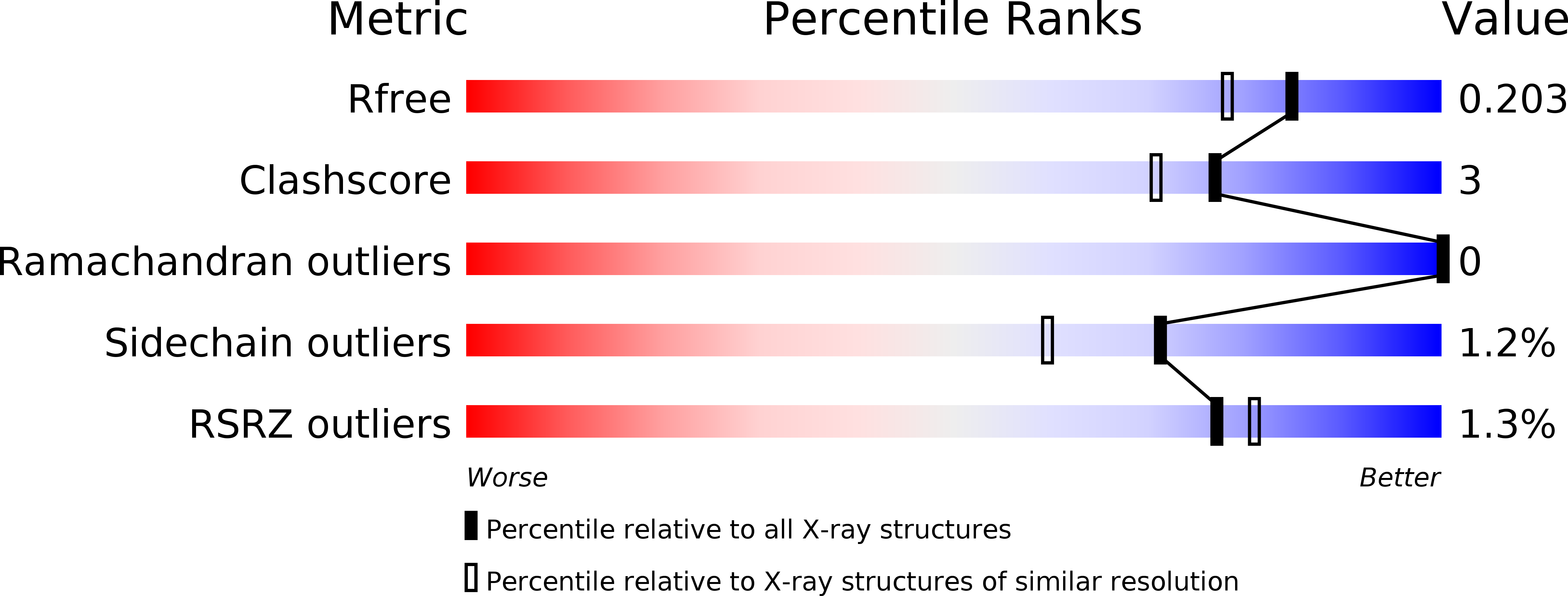
Resolution:
1.70 Å
R-Value Free:
0.19
R-Value Work:
0.15
R-Value Observed:
0.16
Space Group:
P 1 21 1
Macromolecular Entities
Polymer Type:polypeptide(L)
Molecule:SdeD (lpg2509)
Gene (Uniprot):lpg2509
Chain IDs:A
Chain Length:383
Number of Molecules:1
Biological Source:Legionella pneumophila
Polymer Type:polypeptide(L)
Molecule:Polyubiquitin-C
Gene (Uniprot):UBC
Chain IDs:B, C
Chain Length:76
Number of Molecules:2
Biological Source:Homo sapiens
Primary Citation
Mechanism of phosphoribosyl-ubiquitination mediated by a single Legionella effector.
Nature 557 729 733 (2018)
PMID: 29795346 DOI: 10.1038/s41586-018-0147-6

Abstact

Ubiquitination is a post-translational modification that regulates many cellular processes in eukaryotes1-4. The conventional ubiquitination cascade culminates in a covalent linkage between the C terminus of ubiquitin (Ub) and a target protein, usually on a lysine side chain1,5. Recent studies of the Legionella pneumophila SidE family of effector proteins revealed a ubiquitination method in which a phosphoribosyl ubiquitin (PR-Ub) is conjugated to a serine residue on substrates via a phosphodiester bond6-8. Here we present the crystal structure of a fragment of the SidE family member SdeA that retains ubiquitination activity, and determine the mechanism of this unique post-translational modification. The structure reveals that the catalytic module contains two distinct functional units: a phosphodiesterase domain and a mono-ADP-ribosyltransferase domain. Biochemical analysis shows that the mono-ADP-ribosyltransferase domain-mediated conversion of Ub to ADP-ribosylated Ub (ADPR-Ub) and the phosphodiesterase domain-mediated ligation of PR-Ub to substrates are two independent activities of SdeA. Furthermore, we present two crystal structures of a homologous phosphodiesterase domain from the SidE family member SdeD 9 in complexes with Ub and ADPR-Ub. The structures suggest a mechanism for how SdeA processes ADPR-Ub to PR-Ub and AMP, and conjugates PR-Ub to a serine residue in substrates. Our study establishes the molecular mechanism of phosphoribosyl-linked ubiquitination and will enable future studies of this unusual type of ubiquitination in eukaryotes.

Legend

Protein

Chemical

Disease

Primary Citation of related structures