6B67 image
Deposition Date 2017-10-01
Release Date 2018-04-11
Last Version Date 2023-11-15
Entry Detail
PDB ID:
6B67
Keywords:
Title:
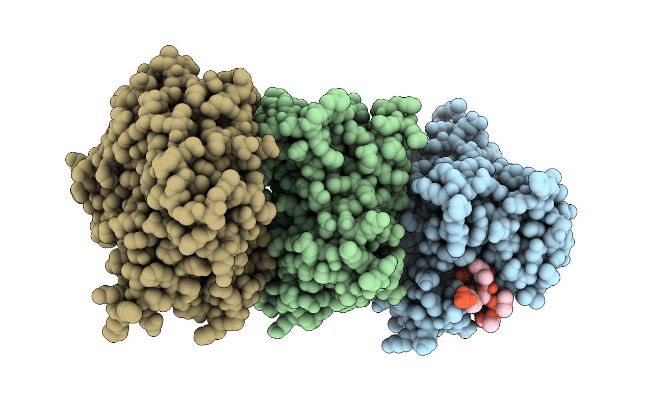
Human PP2Calpha (PPM1A) complexed with cyclic peptide c(MpSIpYVA)
Biological Source:
Source Organism:
Host Organism:
Method Details:
Experimental Method:
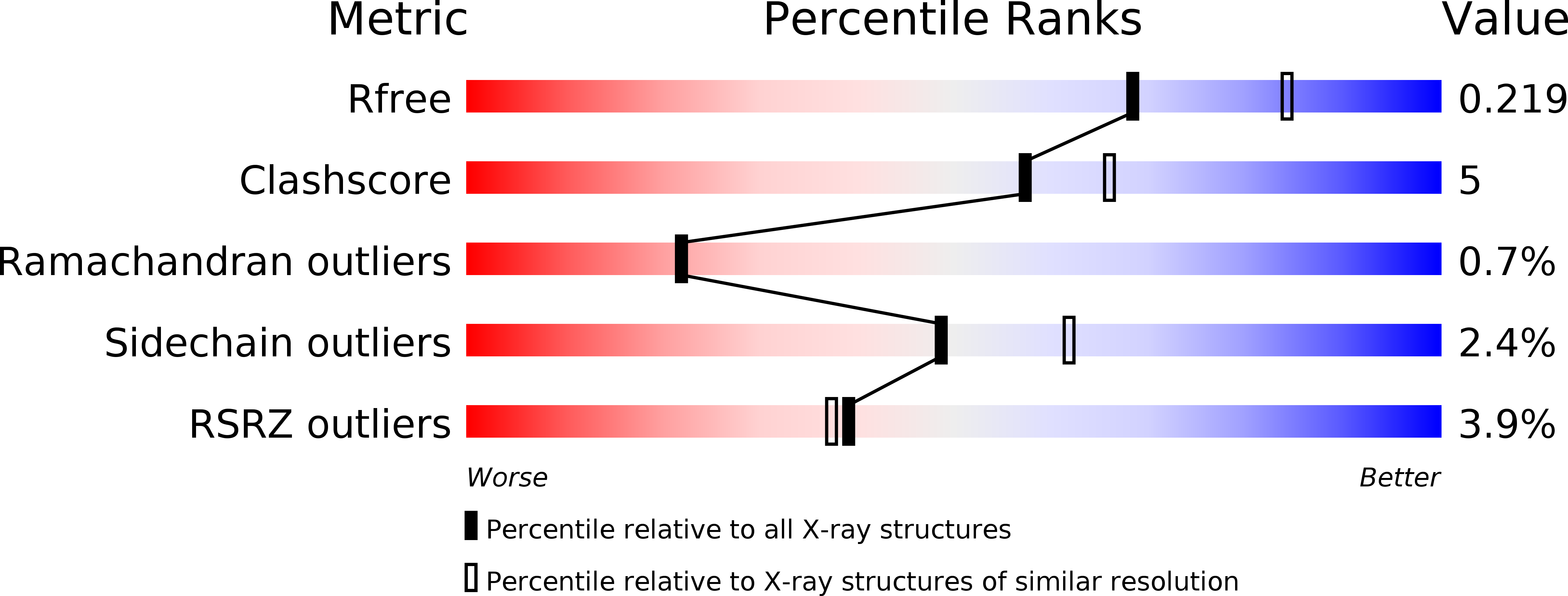
Resolution:
2.20 Å
R-Value Free:
0.22
R-Value Work:
0.16
R-Value Observed:
0.16
Space Group:
C 1 2 1
Macromolecular Entities
Polymer Type:polypeptide(L)
Molecule:Protein phosphatase 1A
Gene (Uniprot):PPM1A
Mutations:D146E
Chain IDs:A, B, C
Chain Length:296
Number of Molecules:3
Biological Source:Homo sapiens
Polymer Type:polypeptide(L)
Molecule:cyclic peptide c(MpSIpYVA)
Chain IDs:D, E, F
Chain Length:7
Number of Molecules:3
Biological Source:synthetic construct
Ligand Molecules
Primary Citation
A trapped human PPM1A-phosphopeptide complex reveals structural features critical for regulation of PPM protein phosphatase activity.
J. Biol. Chem. 293 7993 8008 (2018)
PMID: 29602904 DOI: 10.1074/jbc.RA117.001213

Abstact

Metal-dependent protein phosphatases (PPM) are evolutionarily unrelated to other serine/threonine protein phosphatases and are characterized by their requirement for supplementation with millimolar concentrations of Mg2+ or Mn2+ ions for activity in vitro The crystal structure of human PPM1A (also known as PP2Cα), the first PPM structure determined, displays two tightly bound Mn2+ ions in the active site and a small subdomain, termed the Flap, located adjacent to the active site. Some recent crystal structures of bacterial or plant PPM phosphatases have disclosed two tightly bound metal ions and an additional third metal ion in the active site. Here, the crystal structure of the catalytic domain of human PPM1A, PPM1Acat, complexed with a cyclic phosphopeptide, c(MpSIpYVA), a cyclized variant of the activation loop of p38 MAPK (a physiological substrate of PPM1A), revealed three metal ions in the active site. The PPM1Acat D146E-c(MpSIpYVA) complex confirmed the presence of the anticipated third metal ion in the active site of metazoan PPM phosphatases. Biophysical and computational methods suggested that complex formation results in a slightly more compact solution conformation through reduced conformational flexibility of the Flap subdomain. We also observed that the position of the substrate in the active site allows solvent access to the labile third metal-binding site. Enzyme kinetics of PPM1Acat toward a phosphopeptide substrate supported a random-order, bi-substrate mechanism, with substantial interaction between the bound substrate and the labile metal ion. This work illuminates the structural and thermodynamic basis of an innate mechanism regulating the activity of PPM phosphatases.

Legend

Protein

Chemical

Disease

Primary Citation of related structures