Deposition Date
2017-09-23
Release Date
2017-12-06
Last Version Date
2024-10-16
Entry Detail
PDB ID:
6B3S
Keywords:
Title:
Crystal structure of the Fab fragment of necitumumab (Fab11F8) in complex with domain III from a cetuximab resistant variant of EGFR (sEGFRd3-S468R)
Biological Source:
Source Organism(s):
Homo sapiens (Taxon ID: 9606)
Expression System(s):
Method Details:
Experimental Method:
Resolution:
2.80 Å
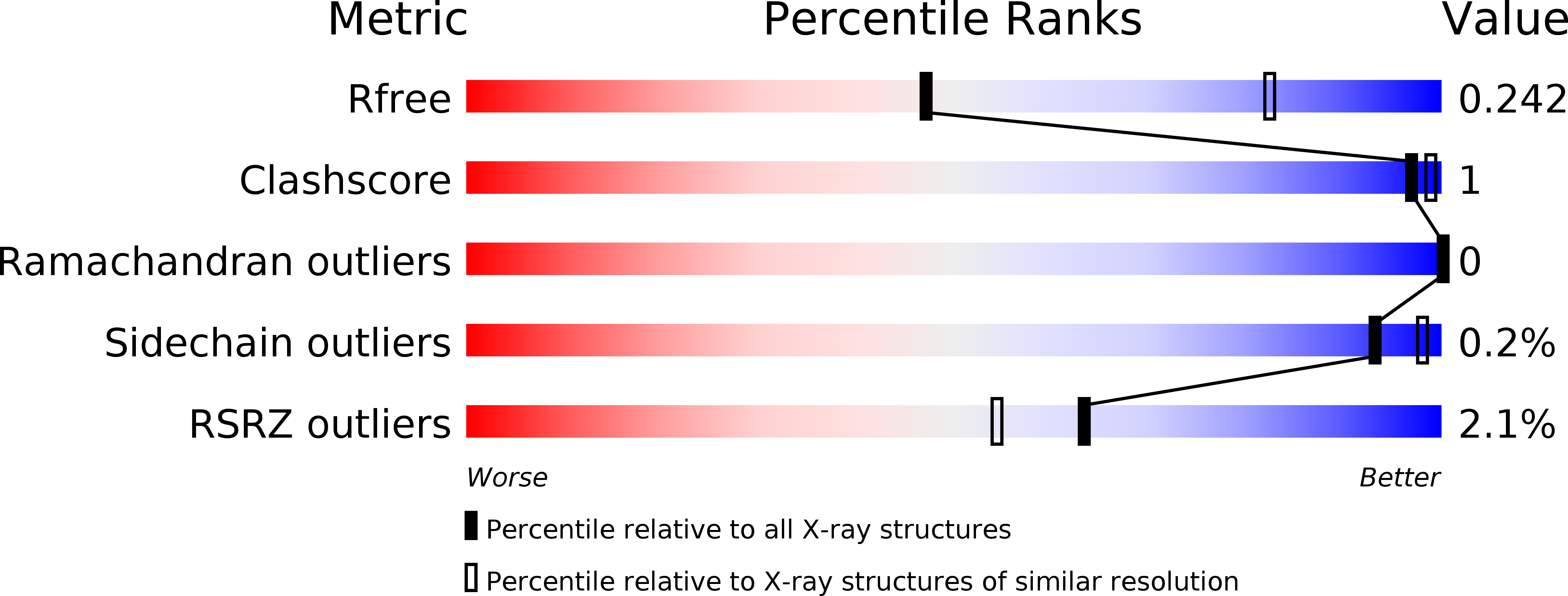
R-Value Free:
0.24
R-Value Work:
0.20
R-Value Observed:
0.20
Space Group:
P 1 2 1