6AYK image
Entry Detail
PDB ID:
6AYK
Keywords:
Title:
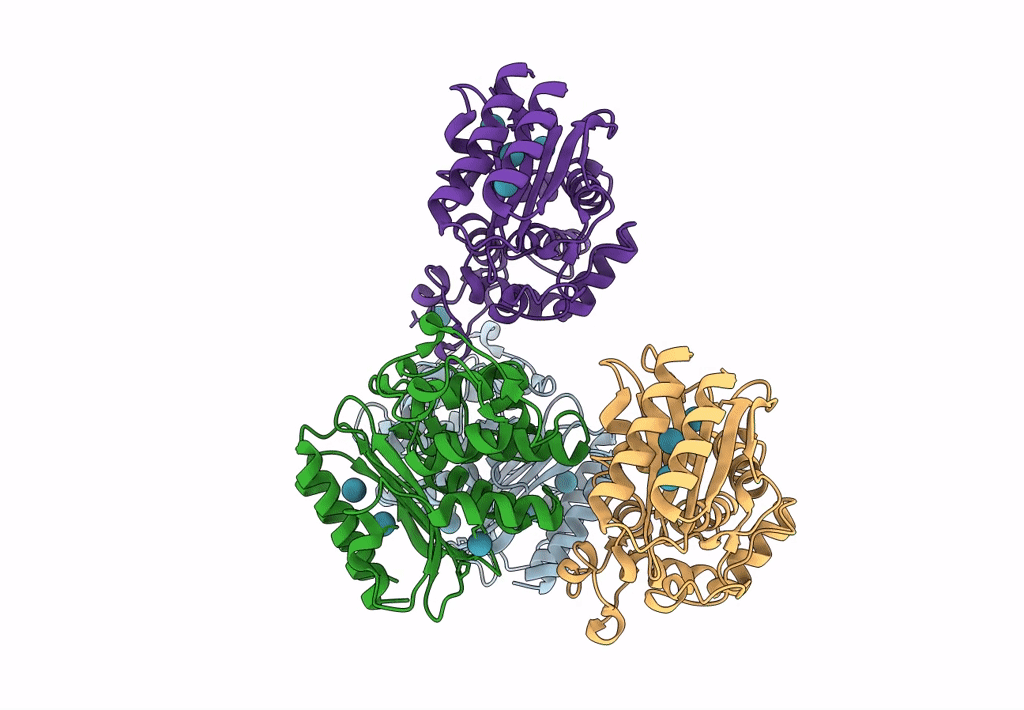
Crystal structure of TEM1 beta-lactamase mutant I263A in the presence of 1.2 MPa xenon
Biological Source:
Source Organism:
PDB Version:
Deposition Date:
2017-09-08
Release Date:
2018-09-12
Method Details:
Experimental Method:
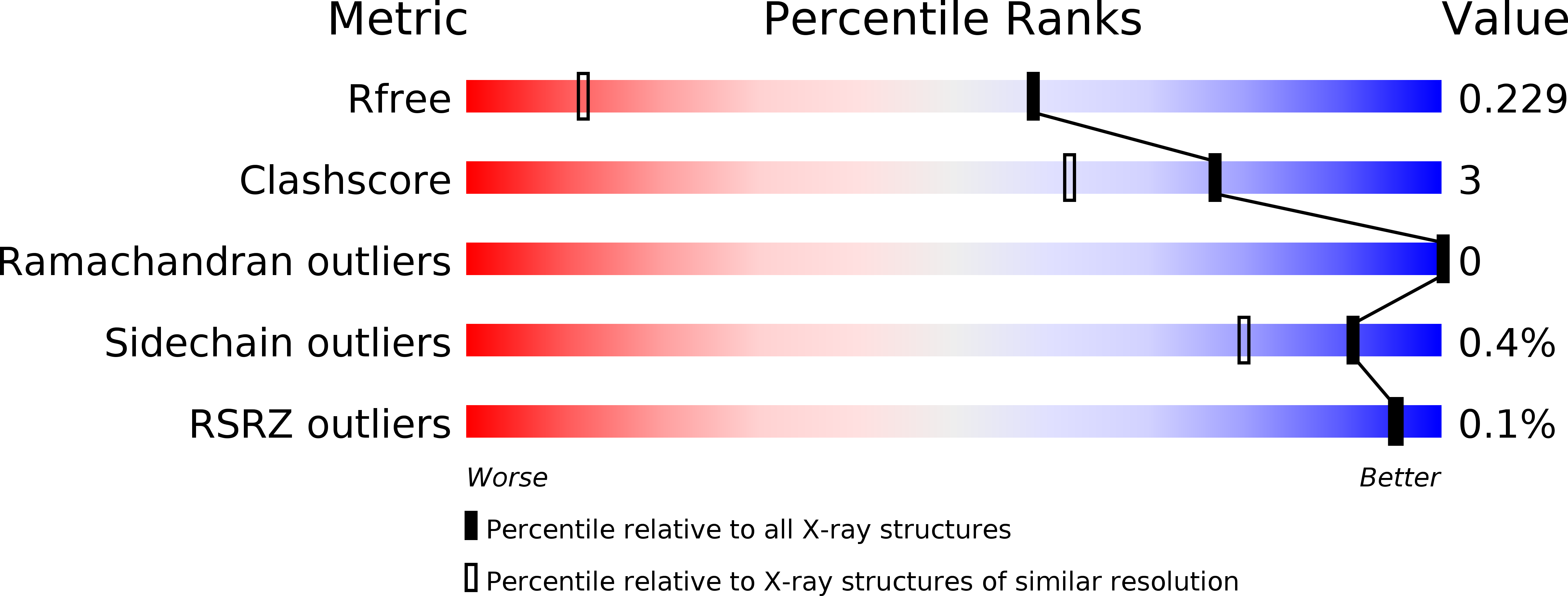
Resolution:
1.44 Å
R-Value Free:
0.21
R-Value Work:
0.19
R-Value Observed:
0.19
Space Group:
P 1 21 1
Macromolecular Entities
Polymer Type:polypeptide(L)
Description:Beta-lactamase TEM
Mutations:M182T, I263A
Chain IDs:A, B, C, D
Chain Length:263
Number of Molecules:4
Biological Source:Escherichia coli
Ligand Molecules
Primary Citation
A Structural Basis for129Xe Hyper-CEST Signal in TEM-1 beta-Lactamase.
Chemphyschem 20 260 267 (2019)
PMID: 30151973 DOI: 10.1002/cphc.201800624

Abstact

Genetically encoded (GE) contrast agents detectable by magnetic resonance imaging (MRI) enable non-invasive visualization of gene expression and cell proliferation at virtually unlimited penetration depths. Using hyperpolarized 129 Xe in combination with chemical exchange saturation transfer, an MR contrast approach known as hyper-CEST, enables ultrasensitive protein detection and biomolecular imaging. GE MRI contrast agents developed to date include nanoscale proteinaceous gas vesicles as well as the monomeric bacterial proteins TEM-1 β-lactamase (bla) and maltose binding protein (MBP). To improve understanding of hyper-CEST NMR with proteins, structural and computational studies were performed to further characterize the Xe-bla interaction. X-ray crystallography validated the location of a high-occupancy Xe binding site predicted by MD simulations, and mutagenesis experiments confirmed this Xe site as the origin of the observed CEST contrast. Structural studies and MD simulations with representative bla mutants offered additional insight regarding the relationship between local protein structure and CEST contrast.

Legend

Protein

Chemical

Disease

Primary Citation of related structures