6AWF image
Entry Detail
PDB ID:
6AWF
Title:
Escherichia coli quinol:fumarate reductase crystallized without dicarboxylate
Biological Source:
Source Organism:
Host Organism:
PDB Version:
Deposition Date:
2017-09-05
Release Date:
2017-12-06
Method Details:
Experimental Method:
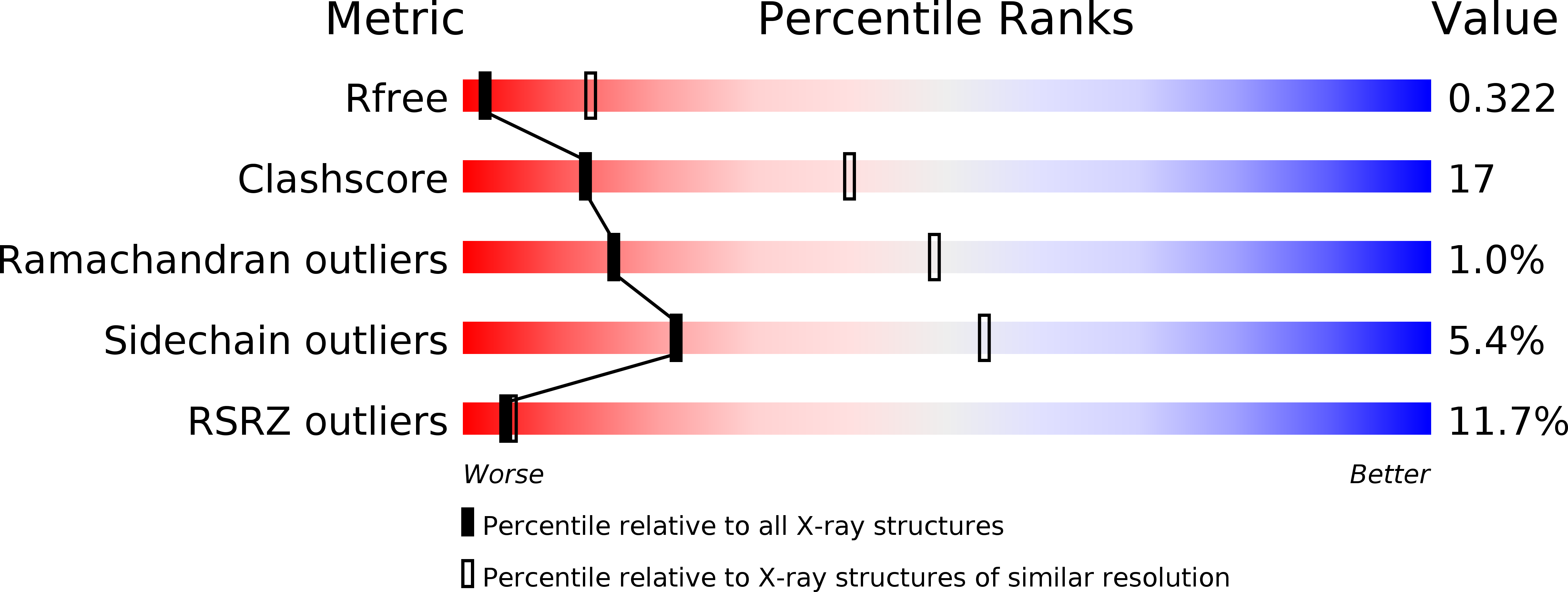
Resolution:
3.35 Å
R-Value Free:
0.31
R-Value Work:
0.28
R-Value Observed:
0.28
Space Group:
P 1 21 1
Macromolecular Entities
Polymer Type:polypeptide(L)
Description:Fumarate reductase flavoprotein subunit
Chain IDs:A, E
Chain Length:602
Number of Molecules:2
Biological Source:Escherichia coli
Polymer Type:polypeptide(L)
Description:Fumarate reductase iron-sulfur subunit
Chain IDs:B, F
Chain Length:243
Number of Molecules:2
Biological Source:Escherichia coli
Polymer Type:polypeptide(L)
Description:Fumarate reductase subunit C
Chain IDs:C, G
Chain Length:130
Number of Molecules:2
Biological Source:Escherichia coli
Polymer Type:polypeptide(L)
Description:Fumarate reductase subunit D
Chain IDs:D, H
Chain Length:119
Number of Molecules:2
Biological Source:Escherichia coli
Primary Citation
New crystal forms of the integral membrane Escherichia coli quinol:fumarate reductase suggest that ligands control domain movement.
J. Struct. Biol. 202 100 104 (2018)
PMID: 29158068 DOI: 10.1016/j.jsb.2017.11.004

Abstact

Quinol:fumarate reductase (QFR) is an integral membrane protein and a member of the respiratory Complex II superfamily. Although the structure of Escherichia coli QFR was first reported almost twenty years ago, many open questions of catalysis remain. Here we report two new crystal forms of QFR, one grown from the lipidic cubic phase and one grown from dodecyl maltoside micelles. QFR crystals grown from the lipid cubic phase processed as P1, merged to 7.5 Å resolution, and exhibited crystal packing similar to previous crystal forms. Crystals grown from dodecyl maltoside micelles processed as P21, merged to 3.35 Å resolution, and displayed a unique crystal packing. This latter crystal form provides the first view of the E. coli QFR active site without a dicarboxylate ligand. Instead, an unidentified anion binds at a shifted position. In one of the molecules in the asymmetric unit, this is accompanied by rotation of the capping domain of the catalytic subunit. In the other molecule, this is associated with loss of interpretable electron density for this same capping domain. Analysis of the structure suggests that the ligand adjusts the position of the capping domain.

Legend

Protein

Chemical

Disease

Primary Citation of related structures