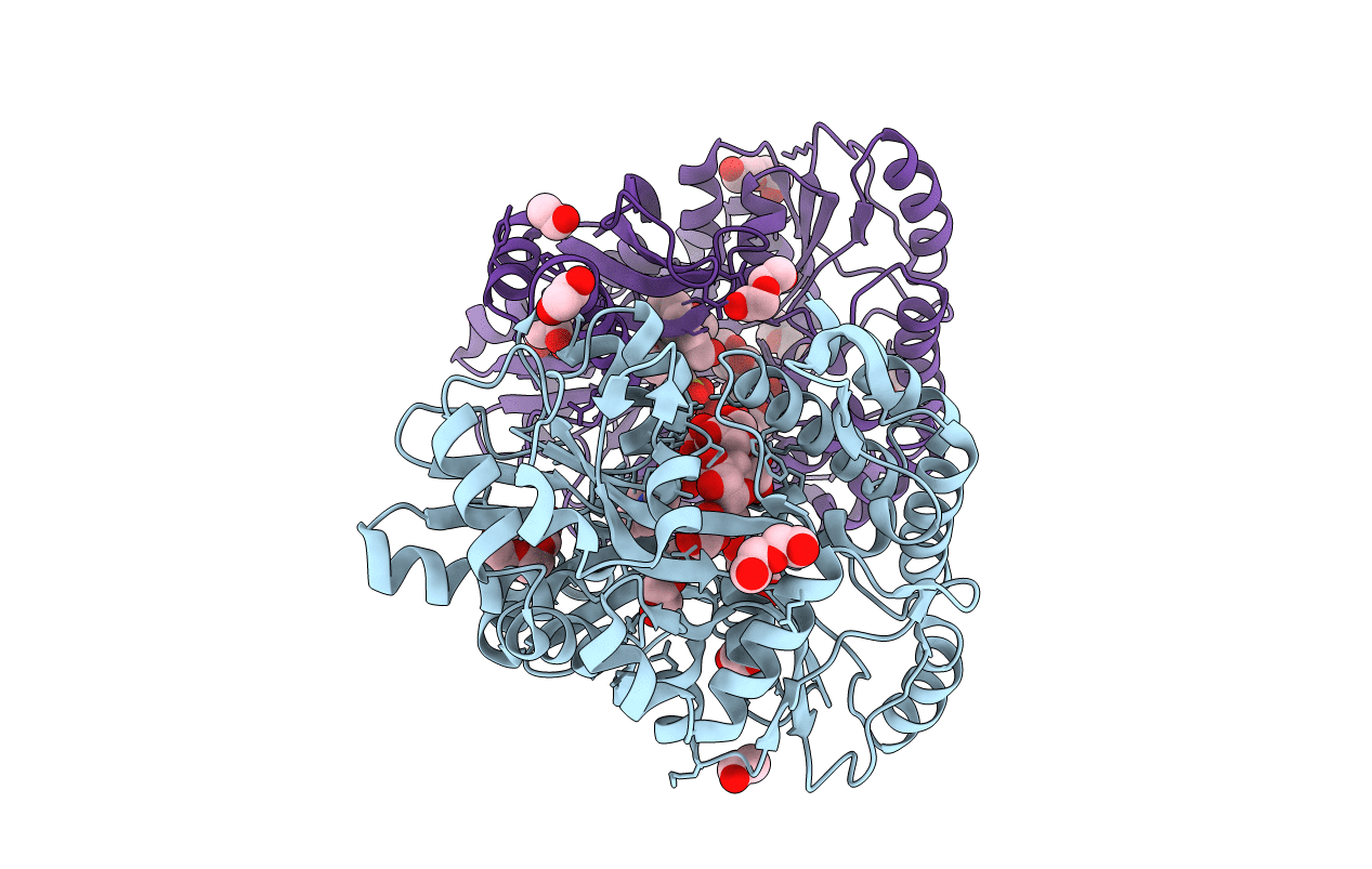
6ANV image
Entry Detail
PDB ID:
6ANV
Keywords:
Title:
Crystal structure of anti-CRISPR protein AcrF1
Biological Source:
PDB Version:
Deposition Date:
2017-08-14
Release Date:
2017-10-25
Method Details:
Experimental Method:
Resolution:
2.27 Å
R-Value Free:
0.22
R-Value Work:
0.16
R-Value Observed:
0.16
Space Group:
P 1 21 1
Macromolecular Entities
Polymer Type:polypeptide(L)
Description:anti-CRISPR protein AcrF1 fused with C-terminal MBP tag
Chain IDs:A, B
Chain Length:452
Number of Molecules:2
Biological Source:Pseudomonas phage JBD30, Escherichia coli O157:H7
Peptide-like Molecules
PRD_900010
Primary Citation
Cryo-EM Structures Reveal Mechanism and Inhibition of DNA Targeting by a CRISPR-Cas Surveillance Complex.
Cell 171 414 426.e12 (2017)
PMID: 28985564 DOI: 10.1016/j.cell.2017.09.006

Abstact

Prokaryotic cells possess CRISPR-mediated adaptive immune systems that protect them from foreign genetic elements, such as invading viruses. A central element of this immune system is an RNA-guided surveillance complex capable of targeting non-self DNA or RNA for degradation in a sequence- and site-specific manner analogous to RNA interference. Although the complexes display considerable diversity in their composition and architecture, many basic mechanisms underlying target recognition and cleavage are highly conserved. Using cryoelectron microscopy (cryo-EM), we show that the binding of target double-stranded DNA (dsDNA) to a type I-F CRISPR system yersinia (Csy) surveillance complex leads to large quaternary and tertiary structural changes in the complex that are likely necessary in the pathway leading to target dsDNA degradation by a trans-acting helicase-nuclease. Comparison of the structure of the surveillance complex before and after dsDNA binding, or in complex with three virally encoded anti-CRISPR suppressors that inhibit dsDNA binding, reveals mechanistic details underlying target recognition and inhibition.

Legend

Protein

Chemical

Disease

Primary Citation of related structures