6AL8 image
Deposition Date 2017-08-07
Release Date 2018-03-07
Last Version Date 2024-03-13
Entry Detail
PDB ID:
6AL8
Keywords:
Title:
Crystal structure HpiC1 Y101F/F138S
Biological Source:
Source Organism:
Host Organism:
Method Details:
Experimental Method:
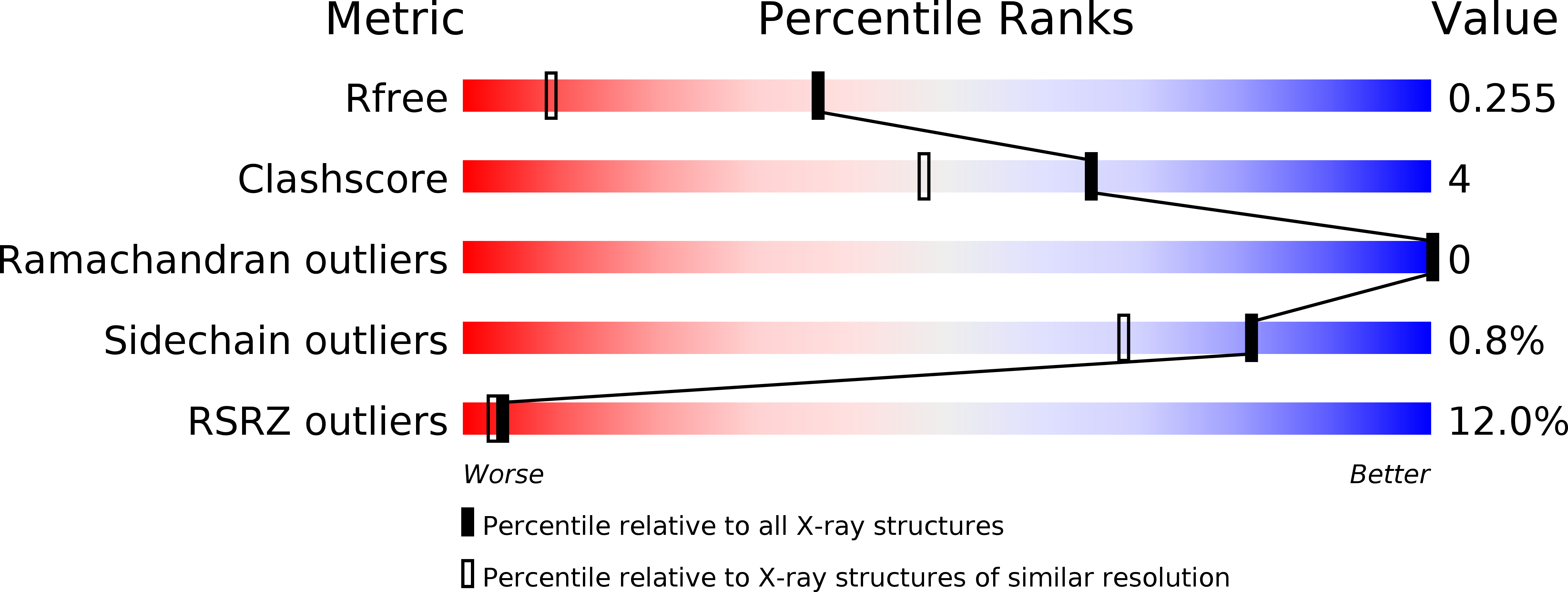
Resolution:
1.64 Å
R-Value Free:
0.25
R-Value Work:
0.22
R-Value Observed:
0.22
Space Group:
P 1 21 1
Macromolecular Entities
Polymer Type:polypeptide(L)
Molecule:12-epi-hapalindole C/U synthase
Gene (Uniprot):hpiU5
Mutations:Y101F, F138S
Chain IDs:A, B, C (auth: D), D (auth: E)
Chain Length:225
Number of Molecules:4
Biological Source:Fischerella sp. ATCC 43239
Primary Citation
Structural basis of the Cope rearrangement and cyclization in hapalindole biogenesis.
Nat. Chem. Biol. 14 345 351 (2018)
PMID: 29531360 DOI: 10.1038/s41589-018-0003-x

Abstact

Hapalindole alkaloids are a structurally diverse class of cyanobacterial natural products defined by their varied polycyclic ring systems and diverse biological activities. These complex metabolites are generated from a common biosynthetic intermediate by the Stig cyclases in three mechanistic steps: a rare Cope rearrangement, 6-exo-trig cyclization, and electrophilic aromatic substitution. Here we report the structure of HpiC1, a Stig cyclase that catalyzes the formation of 12-epi-hapalindole U in vitro. The 1.5-Å structure revealed a dimeric assembly with two calcium ions per monomer and with the active sites located at the distal ends of the protein dimer. Mutational analysis and computational methods uncovered key residues for an acid-catalyzed [3,3]-sigmatropic rearrangement, as well as specific determinants that control the position of terminal electrophilic aromatic substitution, leading to a switch from hapalindole to fischerindole alkaloids.

Legend

Protein

Chemical

Disease

Primary Citation of related structures