6AJD image
Deposition Date 2018-08-27
Release Date 2018-10-24
Last Version Date 2023-11-22
Entry Detail
PDB ID:
6AJD
Keywords:
Title:
Crystal structure of a monometallic dihydropyrimidinase from Pseudomonas aeruginosa PAO1 reveals no lysine carbamylation within the active site
Biological Source:
Method Details:
Experimental Method:
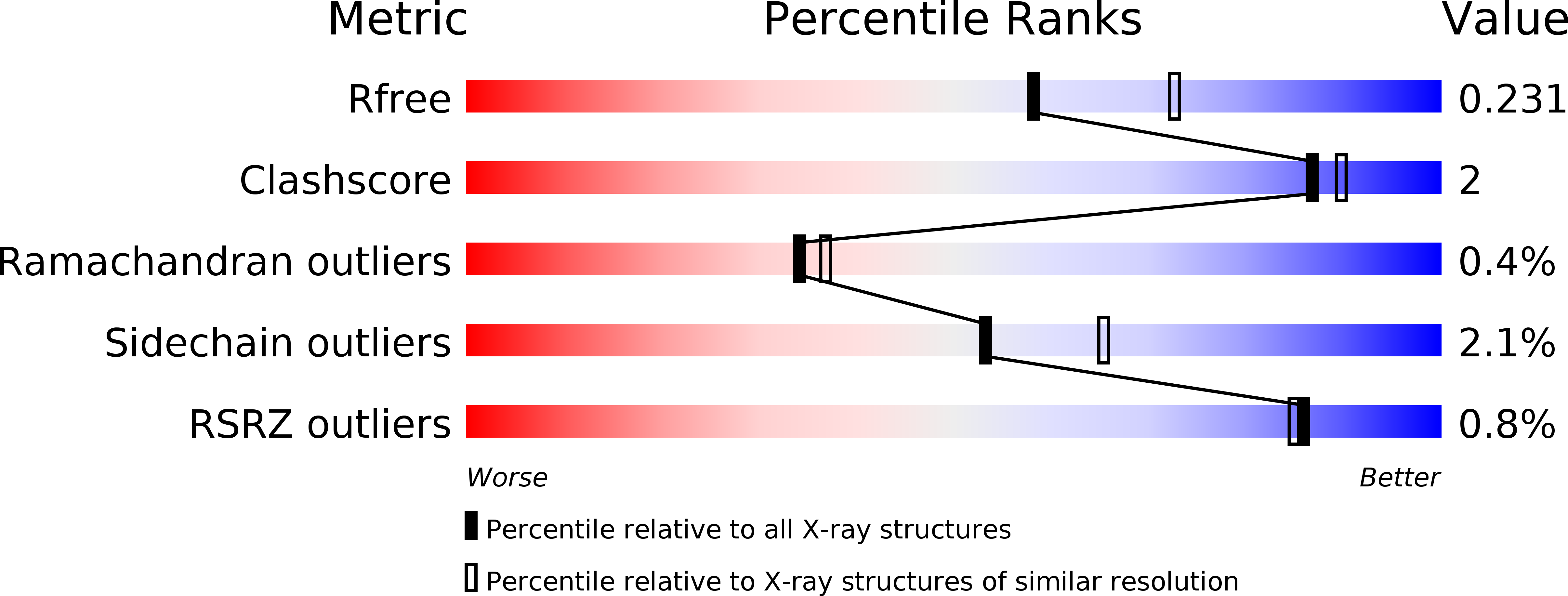
Resolution:
2.22 Å
R-Value Free:
0.23
R-Value Work:
0.18
R-Value Observed:
0.18
Space Group:
P 21 21 2
Macromolecular Entities
Structures with similar UniProt ID
Protein Blast
Polymer Type:polypeptide(L)
Molecule:D-hydantoinase/dihydropyrimidinase
Gene (Uniprot):dht
Chain IDs:A, B
Chain Length:485
Number of Molecules:2
Biological Source:Pseudomonas aeruginosa (strain ATCC 15692 / DSM 22644 / CIP 104116 / JCM 14847 / LMG 12228 / 1C / PRS 101 / PAO1)
Ligand Molecules
Primary Citation
Crystal structures of monometallic dihydropyrimidinase and the human dihydroorotase domain K1556A mutant reveal no lysine carbamylation within the active site
Biochem. Biophys. Res. Commun. 505 439 444 (2018)
PMID: 30268498 DOI: 10.1016/j.bbrc.2018.09.153

Abstact

Dihydropyrimidinase (DHPase) is a member of the cyclic amidohydrolase family, which also includes allantoinase, dihydroorotase (DHOase), hydantoinase, and imidase. Almost all of these zinc metalloenzymes possess a binuclear metal center in which two metal ions are bridged by a post-translational carbamylated Lys. Crystal structure of Tetraodon nigroviridis DHPase reveals that one zinc ion is sufficient to stabilize Lys carbamylation. In this study, we found that one metal coordination was not sufficient to fix CO2 to the Lys in bacterial DHPase. We prepared and characterized mono-Zn DHPase from Pseudomonas aeruginosa (PaDHPase), and the catalytic activity of mono-Zn PaDHPase was not detected. The crystal structure of mono-Zn PaDHPase determined at 2.23 Å resolution (PDB entry 6AJD) revealed that Lys150 was no longer carbamylated. This finding indicated the decarbamylation of the Lys during the metal chelating process. To confirm the state of Lys carbamylation in mono-Zn PaDHPase in solution, mass spectrometric (MS) analysis was carried out. The MS result was in agreement with the theoretical value for uncarbamylated PaDHPase. Crystal structure of the human DHOase domain (huDHOase) K1556A mutant was also determined (PDB entry 5YNZ), and the structure revealed that the active site of huDHOase K1556A mutant contained one metal ion. Like mono-Zn PaDHPase, oxygen ligands of the carbamylated Lys were not required for Znα binding. Considering the collective data from X-ray crystal structure and MS analysis, mono-Zn PaDHPase in both crystalline state and solution was not carbamylated. In addition, structural evidences indicated that post-translational carbamylated Lys was not required for Znα binding in PaDHPase and in huDHOase.

Legend

Protein

Chemical

Disease

Primary Citation of related structures
Feedback Form
Name
Email
Institute
Feedback