Deposition Date
2018-08-20
Release Date
2019-01-23
Last Version Date
2024-03-27
Entry Detail
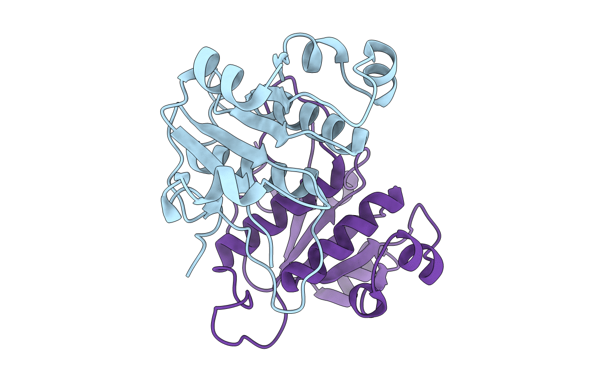
PDB ID:
6AHW
Keywords:
Title:
Crystal structure of circular-permutated YibK methyltransferase from Haemophilus influenzae
Biological Source:
Source Organism(s):
Haemophilus influenzae Rd KW20 (Taxon ID: 71421)
Expression System(s):
Method Details:
Experimental Method:
Resolution:
1.56 Å
R-Value Free:
0.22
R-Value Work:
0.19
R-Value Observed:
0.19
Space Group:
C 1 2 1