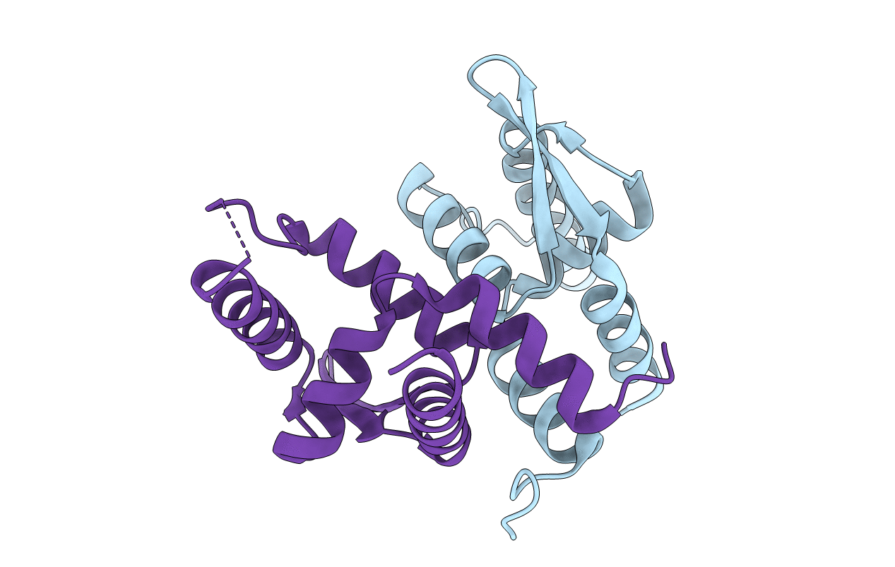
6AHT image
Deposition Date 2018-08-20
Release Date 2019-06-26
Last Version Date 2024-10-30
Entry Detail
PDB ID:
6AHT
Keywords:
Title:
Plasmid partitioning protein TubR from Bacillus cereus
Biological Source:
Source Organism:
Bacillus cereus (Taxon ID: 1396)
Method Details:
Experimental Method:
Resolution:
2.00 Å
R-Value Free:
0.22
R-Value Work:
0.17
Space Group:
P 21 21 21
Macromolecular Entities
Polymer Type:polypeptide(L)
Molecule:Conserved hypothetical plasmid protein
Gene (Uniprot):B4088_5712, BC30052_p1132, BcAH187_pCER270_0087
Chain IDs:A
Chain Length:111
Number of Molecules:1
Biological Source:Bacillus cereus
Polymer Type:polypeptide(L)
Molecule:Conserved hypothetical plasmid protein
Gene (Uniprot):B4088_5712, BC30052_p1132, BcAH187_pCER270_0087
Chain IDs:B
Chain Length:113
Number of Molecules:1
Biological Source:Bacillus cereus
Modified Residue
Compound ID Chain ID Parent Comp ID Details 2D Image
MSE A MET modified residue
Primary Citation
Cooperative DNA Binding of the Plasmid Partitioning Protein TubR from the Bacillus cereus pXO1 Plasmid.
J.Mol.Biol. 430 5015 5028 (2018)
PMID: 30414406 DOI: 10.1016/j.jmb.2018.11.001

Abstact

Tubulin/FtsZ-like GTPase TubZ is responsible for maintaining the stability of pXO1-like plasmids in virulent Bacilli. TubZ forms a filament in a GTP-dependent manner, and like other partitioning systems of low-copy-number plasmids, it requires the centromere-binding protein TubR that connects the plasmid to the TubZ filament. Systems regulating TubZ partitioning have been identified in Clostridium prophages as well as virulent Bacillus species, in which TubZ facilitates partitioning by binding and towing the segrosome: the nucleoprotein complex composed of TubR and the centromere. However, the molecular mechanisms of segrosome assembly and the transient on-off interactions between the segrosome and the TubZ filament remain poorly understood. Here, we determined the crystal structure of TubR from Bacillus cereus at 2.0-Å resolution and investigated the DNA-binding ability of TubR using hydroxyl radical footprinting and electrophoretic mobility shift assays. The TubR dimer possesses 2-fold symmetry and binds to a 15-bp palindromic consensus sequence in the tubRZ promoter region. Continuous TubR-binding sites overlap each other, which enables efficient binding of TubR in a cooperative manner. Interestingly, the segrosome adopts an extended DNA-protein filament structure and likely gains conformational flexibility by introducing non-consensus residues into the palindromes in an asymmetric manner. Together, our experimental results and structural model indicate that the unique centromere recognition mechanism of TubR allows transient complex formation between the segrosome and the dynamic polymer of TubZ.

Legend

Protein

Chemical

Disease

Primary Citation of related structures