6AF6 image
Entry Detail
PDB ID:
6AF6
Title:
PigA with FAD and proline
Biological Source:
Host Organism:
PDB Version:
Deposition Date:
2018-08-08
Release Date:
2018-09-05
Method Details:
Experimental Method:
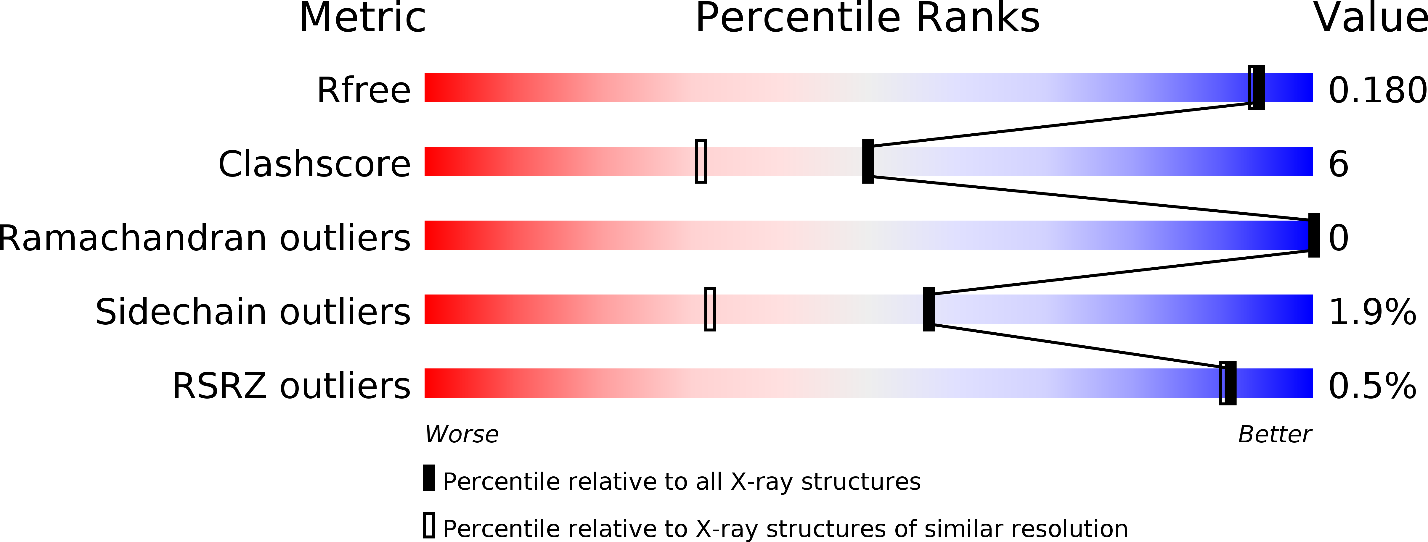
Resolution:
1.62 Å
R-Value Free:
0.17
R-Value Work:
0.15
R-Value Observed:
0.15
Space Group:
I 2 2 2
Macromolecular Entities
Polymer Type:polypeptide(L)
Description:L-prolyl-[peptidyl-carrier protein] dehydrogenase
Mutations:E244A
Chain IDs:A
Chain Length:402
Number of Molecules:1
Biological Source:Serratia sp. (strain ATCC 39006)
Primary Citation
Crystal Structure of PigA: A Prolyl Thioester-Oxidizing Enzyme in Prodigiosin Biosynthesis.
Chembiochem 20 193 202 (2019)
PMID: 30095206 DOI: 10.1002/cbic.201800409

Abstact

Prodigiosin is an intensely red pigment comprising three pyrroles. The biosynthetic pathway includes a two-step proline oxidation catalyzed by phosphatidylinositol N-acetylglucosaminyltransferase subunit A (PigA), with flavin adenine dinucleotide (FAD) as its cofactor. The enzyme is crystallized in the apo form and in complex with FAD and proline. As an acyl coenzyme A dehydrogenase (ACAD) family member, the protein folds into a β-sheet flanked by two α-helical domains. PigA forms a tetramer, which is consistent with analytical ultracentrifugation results. FAD binds to PigA in a similar way to that in the other enzymes of the ACAD family. The variable conformations of loop β4-β5 and helix αG correlate well with the structural flexibility required for substrate entrance to the Re side of FAD. Modeling with PigG, the acyl carrier protein, suggests a reasonable mode of interaction with PigA. The structure helps to explain the proline oxidation mechanism, in which Glu244 plays a central role by abstracting the substrate protons. It also reveals a plausible pocket for oxygen binding to the Si side of FAD.

Legend

Protein

Chemical

Disease

Primary Citation of related structures