6AEM image
Deposition Date 2018-08-05
Release Date 2018-12-12
Last Version Date 2023-11-22
Entry Detail
PDB ID:
6AEM
Title:
Crystal structure of the PKD1 domain of Vibrio anguillarum Epp
Biological Source:
Source Organism:
Host Organism:
Method Details:
Experimental Method:
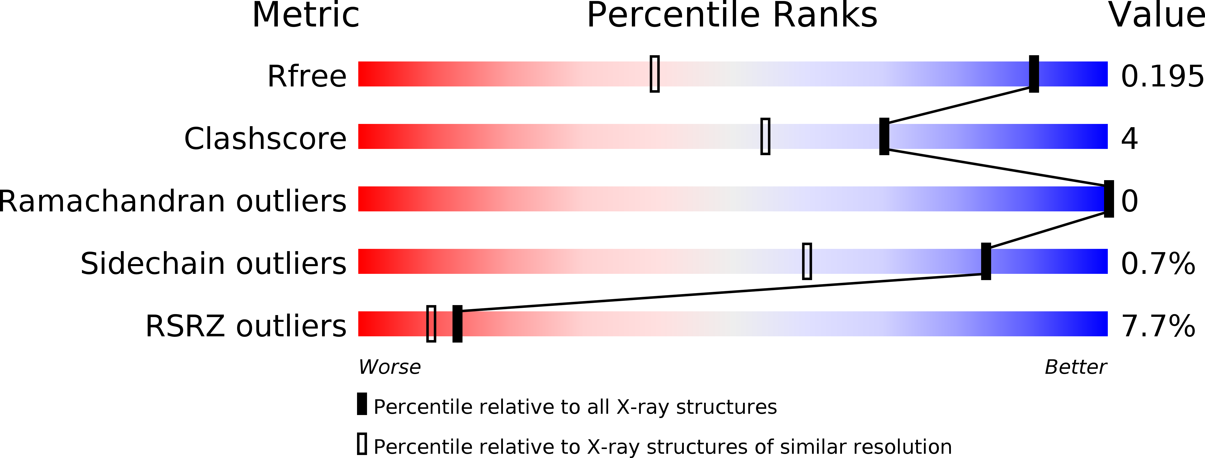
Resolution:
1.27 Å
R-Value Free:
0.18
R-Value Work:
0.15
R-Value Observed:
0.15
Space Group:
C 1 2 1
Macromolecular Entities
Structures with similar UniProt ID
Protein Blast
Polymer Type:polypeptide(L)
Molecule:PKD domain
Chain IDs:A, B
Chain Length:87
Number of Molecules:2
Biological Source:Vibrio anguillarum
Primary Citation
Structural basis for specific calcium binding by the polycystic-kidney-disease domain of Vibrio anguillarum protease Epp
Biochem. Biophys. Res. Commun. 505 471 477 (2018)
PMID: 30268503 DOI: 10.1016/j.bbrc.2018.09.108

Abstact

Extracellular proteases are often produced as pre-pro-enzyme and then undergo multiple processing steps to mature into the active form. The protease Epp, a virulent factor of Vibrio anguillarum, belongs to this family. Its maturation might be regulated by Ca2+ via its polycystic kidney disease (PKD) domain, but the molecular mechanism is unknown. Herein, we report the crystal structure of the first PKD domain from V. anguillarum Epp (Epp-PKD1) and its specific Ca2+-binding capacity. Epp-PKD1 exists as a monomer, consisting of seven β-strands which form two β-sheets stacking with each other. One Ca2+ is bound by the residues Asn3, Gln4, Asp27, Asp29, Asp68 and a water molecule with a pentagonal bipyramidal geometry. Incubating the apo Epp-PKD1 with Ca2+ but not Mg2+, Mn2+, or Zn2+, enhances the thermal and chemical stability of Epp-PKD1, indicating its specific binding to Ca2+. Epp-PKD1 shares high similarity in both sequence and overall structure with that of Vibrio cholerae PrtV, a homologous protease of Epp, however, they differ in the oligomeric state and local structure at the Ca2+-binding site, suggesting maturation of PrtV and Epp might be differently regulated by Ca2+. Likely, proteases may take advantage of the structural diversity in PKD domains to tune their Ca2+-regulated maturation process.

Legend

Protein

Chemical

Disease

Primary Citation of related structures
Feedback Form
Name
Email
Institute
Feedback