6AD9 image
Deposition Date 2018-07-31
Release Date 2018-11-14
Last Version Date 2024-03-27
Entry Detail
PDB ID:
6AD9
Keywords:
Title:
Crystal Structure of PPARgamma Ligand Binding Domain in complex with dibenzooxepine derivative compound-9
Biological Source:
Source Organism:
Homo sapiens (Taxon ID: 9606)
Host Organism:
Method Details:
Experimental Method:
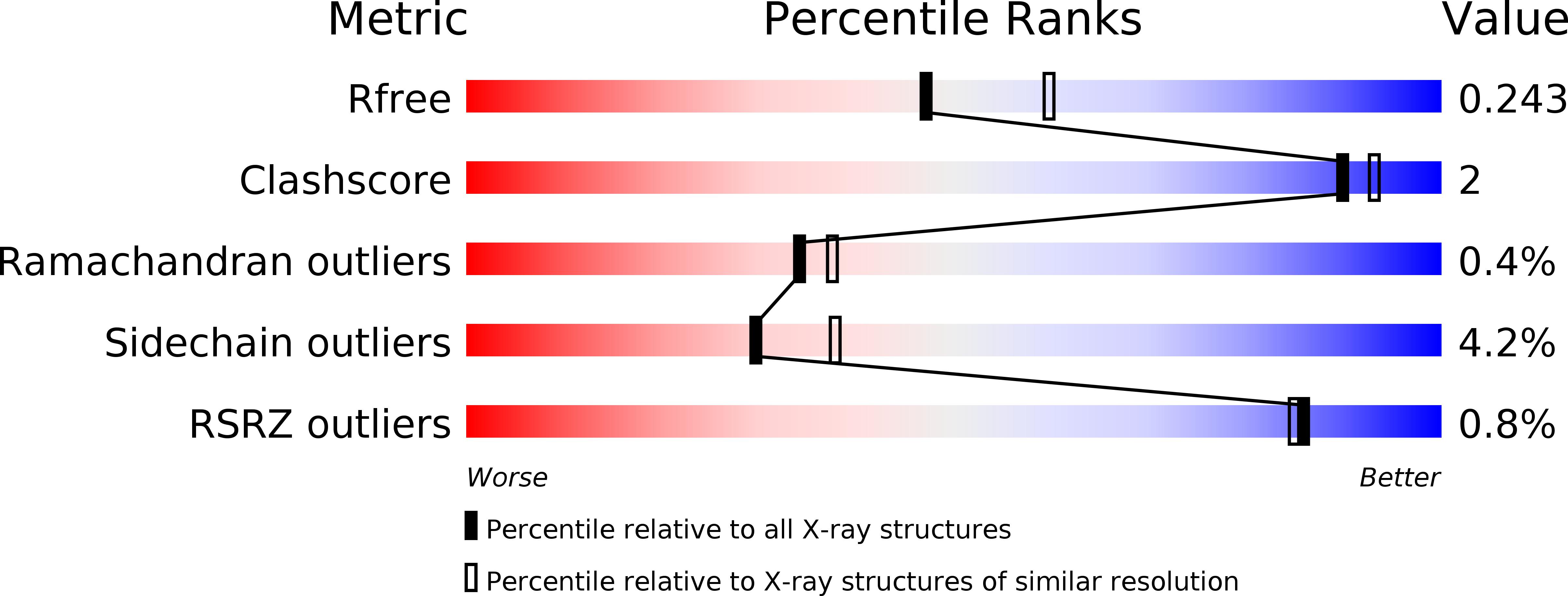
Resolution:
2.20 Å
R-Value Free:
0.23
R-Value Work:
0.18
R-Value Observed:
0.18
Space Group:
P 1 21 1
Macromolecular Entities
Polymer Type:polypeptide(L)
Molecule:Peroxisome proliferator-activated receptor gamma
Gene (Uniprot):PPARG
Chain IDs:A
Chain Length:287
Number of Molecules:1
Biological Source:Homo sapiens
Polymer Type:polypeptide(L)
Molecule:12-mer peptide from Peroxisome proliferator-activated receptor gamma coactivator 1-alpha
Gene (Uniprot):PPARGC1A
Chain IDs:B
Chain Length:12
Number of Molecules:1
Biological Source:Homo sapiens
Ligand Molecules
Primary Citation
Development of Dihydrodibenzooxepine Peroxisome Proliferator-Activated Receptor (PPAR) Gamma Ligands of a Novel Binding Mode as Anticancer Agents: Effective Mimicry of Chiral Structures by Olefinic E/ Z-Isomers.
J. Med. Chem. 61 10067 10083 (2018)
PMID: 30351933 DOI: 10.1021/acs.jmedchem.8b01200

Abstact

A novel class of PPARγ ligand 1 (EC50 = 197 nM) with a dibenzoazepin scaffold was identified through high-throughput screening campaign. To avoid the synthetically troublesome chiral center of 1, its conformational analysis using the MacroModel was conducted, focusing on conformational flip of the tricyclic ring and the conformational restriction by the methyl group at the chiral center. On the basis of this analysis, scaffold hopping of dibenzoazepine into dibenzo[ b, e]oxepine by replacing the chiral structures with the corresponding olefinic E/ Z isomers was performed. Consequently, dibenzo[ b, e]oxepine scaffold 9 was developed showing extremely potent PPARγ reporter activity (EC50 = 2.4 nM, efficacy = 9.5%) as well as differentiation-inducing activity against a gastric cancer cell line MKN-45 that was more potent than any other well-known PPARγ agonists in vitro (94% at 30 nM). The X-ray crystal structure analysis of 9 complexed with PPARγ showed that it had a unique binding mode to PPARγ ligand-binding domain that differed from that of any other PPARγ agonists identified thus far.

Legend

Protein

Chemical

Disease

Primary Citation of related structures