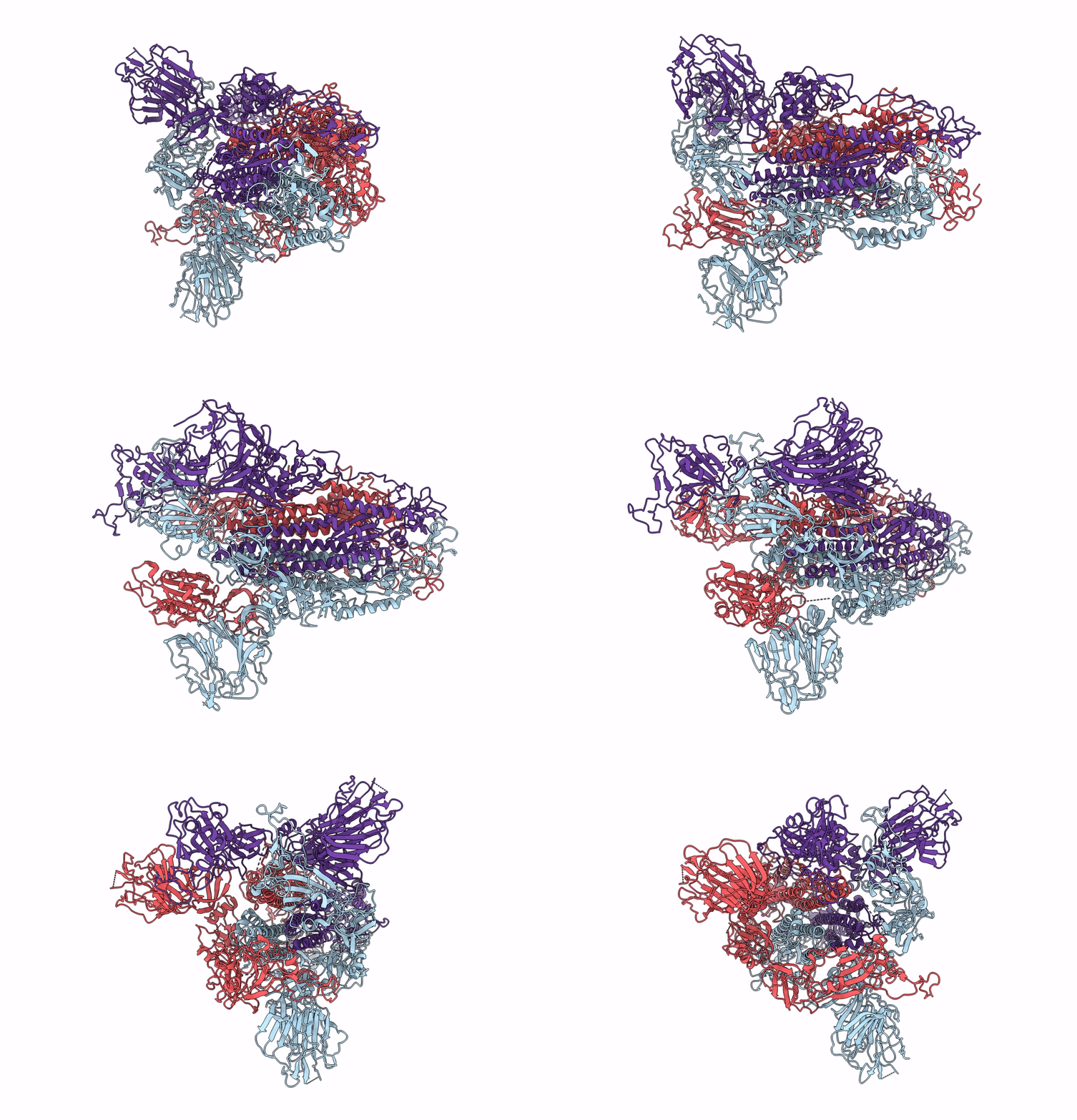
6ACD image
Deposition Date 2018-07-26
Release Date 2018-08-08
Last Version Date 2024-10-23
Entry Detail
PDB ID:
6ACD
Keywords:
Title:
Trypsin-cleaved and low pH-treated SARS-CoV spike glycoprotein and ACE2 complex, ACE2-free conformation with one RBD in up conformation
Biological Source:
Source Organism:
Host Organism:
Method Details:
Experimental Method:
Resolution:
3.90 Å
Aggregation State:
PARTICLE
Reconstruction Method:
SINGLE PARTICLE
Macromolecular Entities
Polymer Type:polypeptide(L)
Molecule:Spike glycoprotein
Gene (Uniprot):S
Chain IDs:A, B, C
Chain Length:1203
Number of Molecules:3
Biological Source:Human SARS coronavirus
Ligand Molecules
Primary Citation
Cryo-EM structure of the SARS coronavirus spike glycoprotein in complex with its host cell receptor ACE2.
PLoS Pathog. 14 e1007236 e1007236 (2018)
PMID: 30102747 DOI: 10.1371/journal.ppat.1007236

Abstact

The trimeric SARS coronavirus (SARS-CoV) surface spike (S) glycoprotein consisting of three S1-S2 heterodimers binds the cellular receptor angiotensin-converting enzyme 2 (ACE2) and mediates fusion of the viral and cellular membranes through a pre- to postfusion conformation transition. Here, we report the structure of the SARS-CoV S glycoprotein in complex with its host cell receptor ACE2 revealed by cryo-electron microscopy (cryo-EM). The complex structure shows that only one receptor-binding domain of the trimeric S glycoprotein binds ACE2 and adopts a protruding "up" conformation. In addition, we studied the structures of the SARS-CoV S glycoprotein and its complexes with ACE2 in different in vitro conditions, which may mimic different conformational states of the S glycoprotein during virus entry. Disassociation of the S1-ACE2 complex from some of the prefusion spikes was observed and characterized. We also characterized the rosette-like structures of the clustered SARS-CoV S2 trimers in the postfusion state observed on electron micrographs. Structural comparisons suggested that the SARS-CoV S glycoprotein retains a prefusion architecture after trypsin cleavage into the S1 and S2 subunits and acidic pH treatment. However, binding to the receptor opens up the receptor-binding domain of S1, which could promote the release of the S1-ACE2 complex and S1 monomers from the prefusion spike and trigger the pre- to postfusion conformational transition.

Legend

Protein

Chemical

Disease

Primary Citation of related structures