6AC5 image
Deposition Date 2018-07-25
Release Date 2019-05-01
Last Version Date 2024-10-23
Entry Detail
PDB ID:
6AC5
Title:
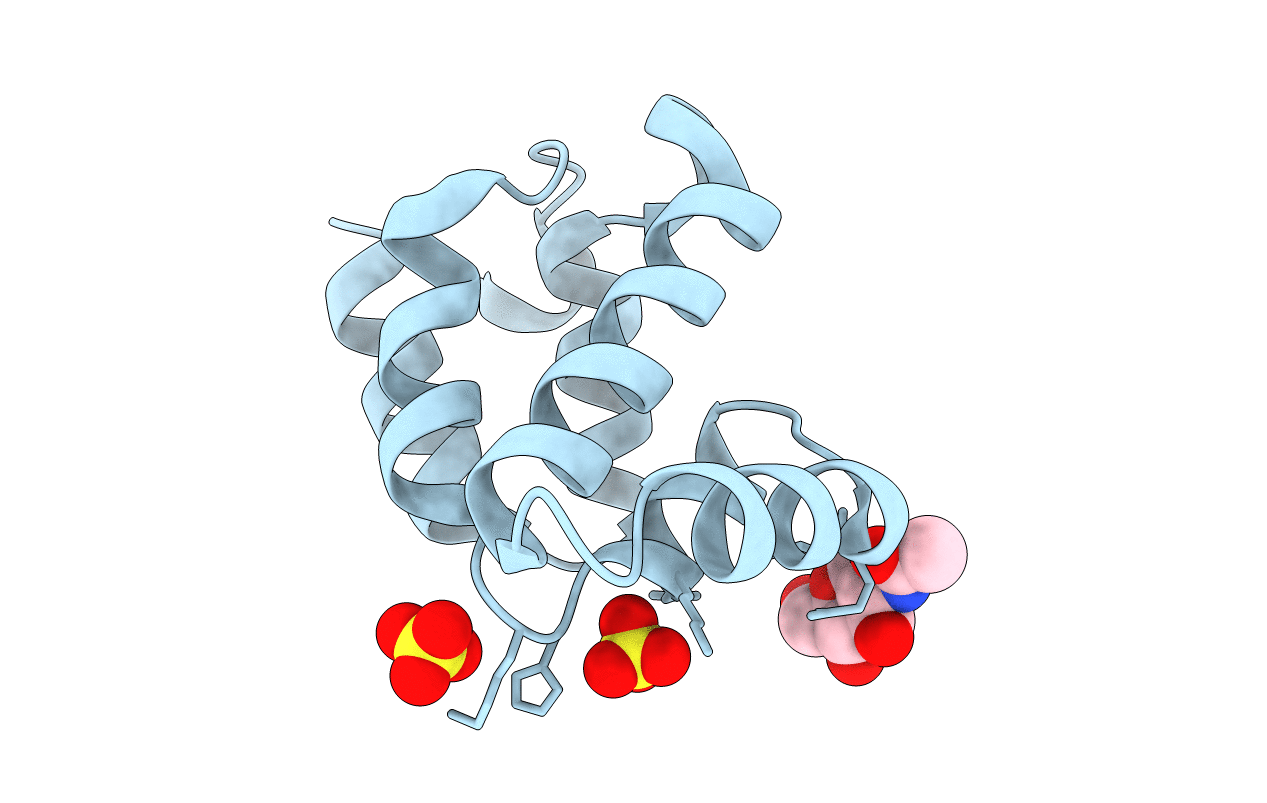
Crystal structure of RIPK1 death domain GlcNAcylated by EPEC effector NleB
Biological Source:
Source Organism:
Homo sapiens (Taxon ID: 9606)
Method Details:
Experimental Method:
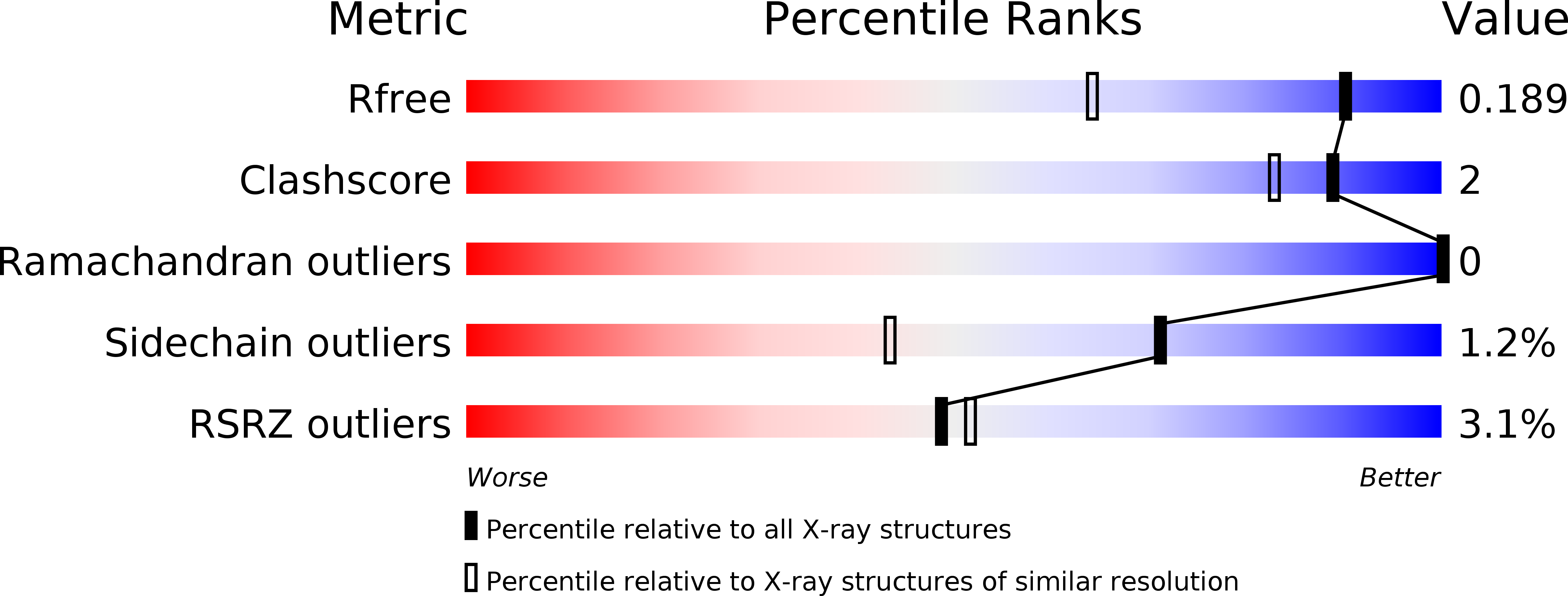
Resolution:
1.45 Å
R-Value Free:
0.18
R-Value Work:
0.15
R-Value Observed:
0.15
Space Group:
P 43 21 2
Macromolecular Entities
Polymer Type:polypeptide(L)
Molecule:Receptor-interacting serine/threonine-protein kinase 1
Gene (Uniprot):RIPK1
Chain IDs:A
Chain Length:111
Number of Molecules:1
Biological Source:Homo sapiens
Primary Citation
Structural and Functional Insights into Host Death Domains Inactivation by the Bacterial Arginine GlcNAcyltransferase Effector.
Mol.Cell 74 922 ? (2019)
PMID: 30979585 DOI: 10.1016/j.molcel.2019.03.028

Abstact

Enteropathogenic E. coli NleB and related type III effectors catalyze arginine GlcNAcylation of death domain (DD) proteins to block host defense, but the underlying mechanism is unknown. Here we solve crystal structures of NleB alone and in complex with FADD-DD, UDP, and Mn2+ as well as NleB-GlcNAcylated DDs of TRADD and RIPK1. NleB adopts a GT-A fold with a unique helix-pair insertion to hold FADD-DD; the interface contacts explain the selectivity of NleB for certain DDs. The acceptor arginine is fixed into a cleft, in which Glu253 serves as a base to activate the guanidinium. Analyses of the enzyme-substrate complex and the product structures reveal an inverting sugar-transfer reaction and a detailed catalytic mechanism. These structural insights are validated by mutagenesis analyses of NleB-mediated GlcNAcylation in vitro and its function in mouse infection. Our study builds a structural framework for understanding of NleB-catalyzed arginine GlcNAcylation of host death domain.

Legend

Protein

Chemical

Disease

Primary Citation of related structures