Deposition Date
2018-07-18
Release Date
2018-08-15
Last Version Date
2024-11-13
Entry Detail
PDB ID:
6AAH
Keywords:
Title:
Crystal structure of JAK1 in complex with peficitinib
Biological Source:
Source Organism(s):
Homo sapiens (Taxon ID: 9606)
Expression System(s):
Method Details:
Experimental Method:
Resolution:
1.83 Å
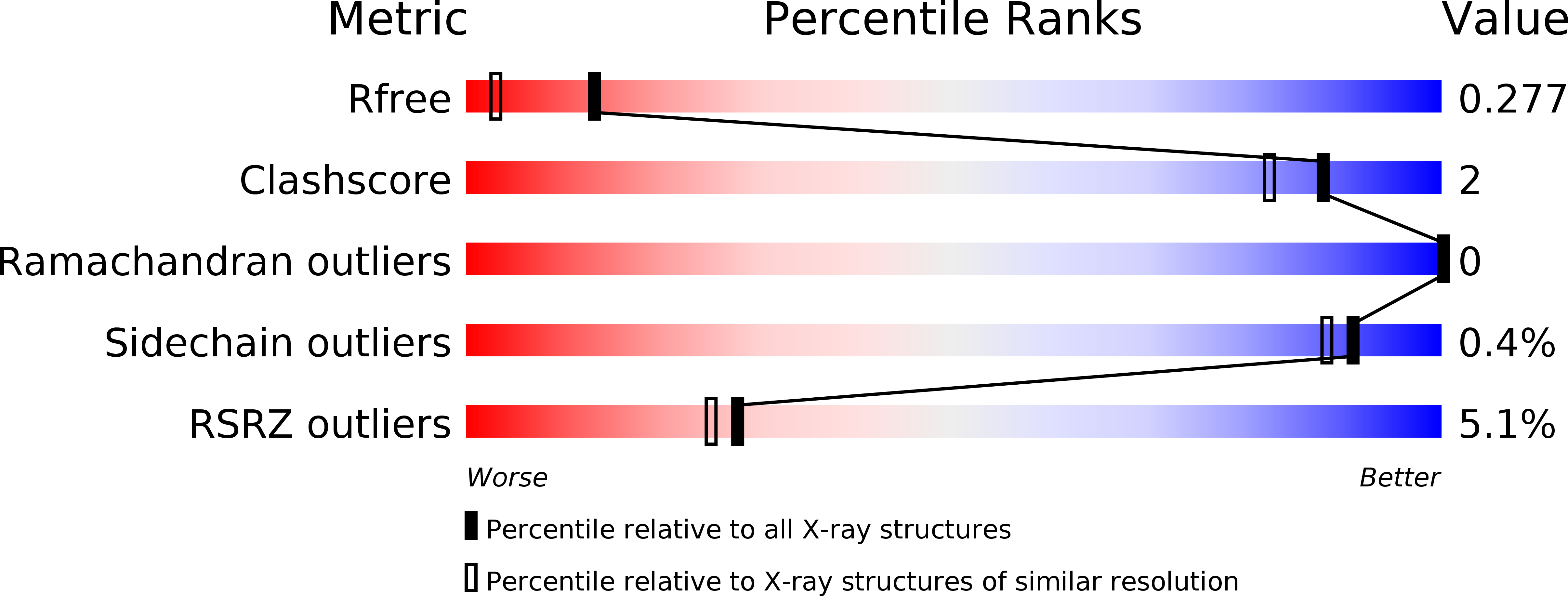
R-Value Free:
0.25
R-Value Work:
0.22
R-Value Observed:
0.22
Space Group:
P 21 21 21