6AAA image
Deposition Date 2018-07-18
Release Date 2018-08-22
Last Version Date 2023-11-22
Entry Detail
PDB ID:
6AAA
Title:
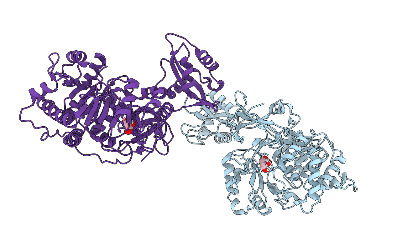
Structure of a blue-shifted Luciferase from Amydetes vivianii
Biological Source:
Source Organism:
Amydetes (Taxon ID: 1176510)
Host Organism:
Method Details:
Experimental Method:
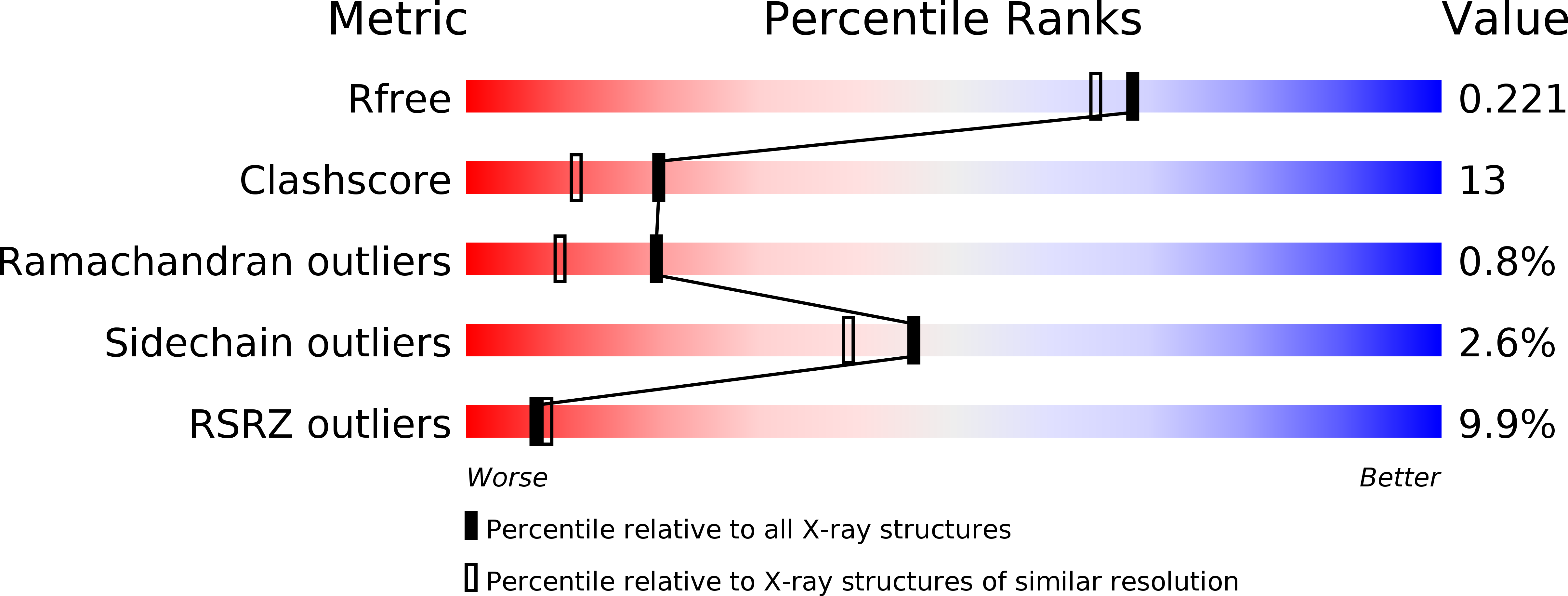
Resolution:
1.90 Å
R-Value Free:
0.21
R-Value Work:
0.19
R-Value Observed:
0.19
Space Group:
P 21 21 21
Macromolecular Entities
Structures with similar UniProt ID
Protein Blast
Polymer Type:polypeptide(L)
Molecule:Blue-shifted Luciferase
Chain IDs:A, B
Chain Length:547
Number of Molecules:2
Biological Source:Amydetes
Ligand Molecules
Primary Citation
Beetle luciferases with naturally red- and blue-shifted emission.
Life Sci Alliance 1 e201800072 e201800072 (2018)
PMID: 30456363 DOI: 10.26508/lsa.201800072

Abstact

The different colors of light emitted by bioluminescent beetles that use an identical substrate and chemiexcitation reaction sequence to generate light remain a challenging and controversial mechanistic conundrum. The crystal structures of two beetle luciferases with red- and blue-shifted light relative to the green yellow light of the common firefly species provide direct insight into the molecular origin of the bioluminescence color. The structure of a blue-shifted green-emitting luciferase from the firefly Amydetes vivianii is monomeric with a structural fold similar to the previously reported firefly luciferases. The only known naturally red-emitting luciferase from the glow-worm Phrixothrix hirtus exists as tetramers and octamers. Structural and computational analyses reveal varying aperture between the two domains enclosing the active site. Mutagenesis analysis identified two conserved loops that contribute to the color of the emitted light. These results are expected to advance comparative computational studies into the conformational landscape of the luciferase reaction sequence.

Legend

Protein

Chemical

Disease

Primary Citation of related structures