6A9W image
Deposition Date 2018-07-16
Release Date 2019-03-13
Last Version Date 2024-03-27
Entry Detail
PDB ID:
6A9W
Keywords:
Title:
Structure of the bifunctional DNA primase-polymerase from phage NrS-1
Biological Source:
Source Organism:
Host Organism:
Method Details:
Experimental Method:
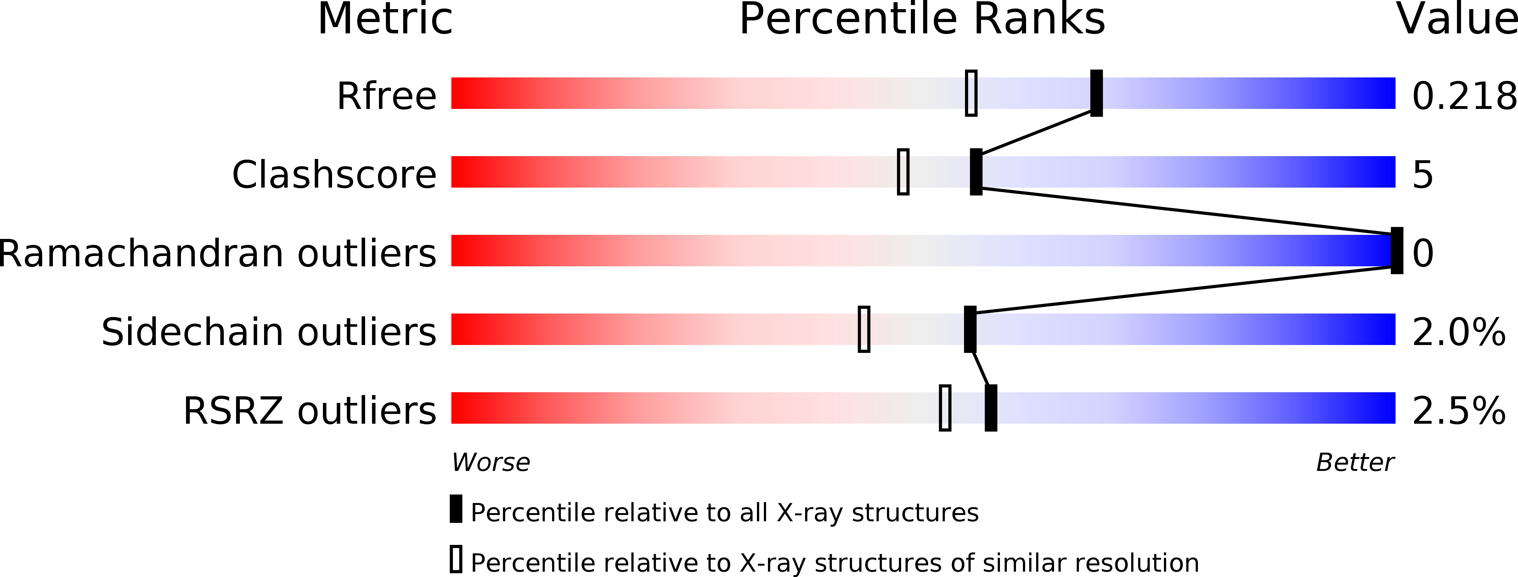
Resolution:
1.80 Å
R-Value Free:
0.21
R-Value Work:
0.17
R-Value Observed:
0.18
Space Group:
C 1 2 1
Macromolecular Entities
Structures with similar UniProt ID
Protein Blast
Polymer Type:polypeptide(L)
Molecule:Primase
Gene (Uniprot):28
Chain IDs:A
Chain Length:320
Number of Molecules:1
Biological Source:Nitratiruptor phage NrS-1
Primary Citation
Crystal structure and biochemical studies of the bifunctional DNA primase-polymerase from phage NrS-1.
Biochem. Biophys. Res. Commun. 510 573 579 (2019)
PMID: 30739783 DOI: 10.1016/j.bbrc.2019.01.144

Abstact

A novel DNA polymerase found in the deep-sea vent phage NrS-1, was confirmed to have both DNA polymerase and primase activities. In this polymerase, the N-terminal residues 1-300 (referred to as N300) are the core region required for polymerizing DNA and catalyzing de novo DNA synthesis. Here, the crystal structure of N300 was solved at a resolution of 1.80 Å. The overall structure consists of a prim/pol domain and a helix bundle domain, which are separated by a 14-residue-long flexible tether (residues 177-190). Both the prim/pol domain of N300 and other primase-polymerases (prim-pol) encompass an analogous fold with conserved catalytic residues. Mutagenesis and enzymatic activity assays show that the acidic active-site residue E139 is required for both polymerase and primase activities. Functional assays confirm the essentiality of the helix bundle domain for primase activity. Furthermore, we identified a mutant (N300-Y261A) of the helix bundle domain, which probably plays an indispensable role in the primer initiation and recognition of template DNA.

Legend

Protein

Chemical

Disease

Primary Citation of related structures