
Deposition Date
2018-07-13
Release Date
2019-06-12
Last Version Date
2023-11-22
Entry Detail
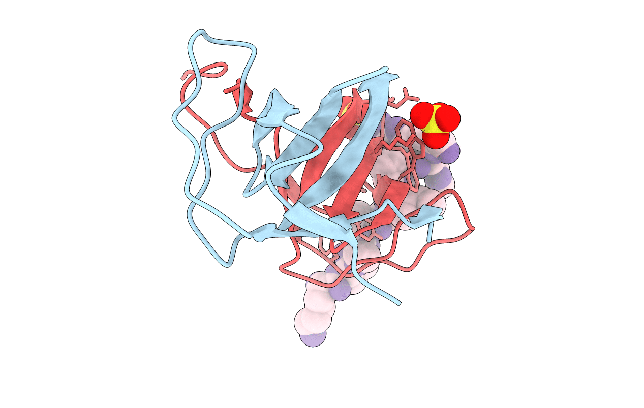
PDB ID:
6A9C
Keywords:
Title:
Crystal Structure c-terminal SH3 domain of Myosin IB from Entamoeba histolytica bound to EhFP10(GEF) peptide.
Biological Source:
Source Organism(s):
Entamoeba histolytica (Taxon ID: 5759)
Expression System(s):
Method Details:
Experimental Method:
Resolution:
1.98 Å
R-Value Free:
0.24
R-Value Work:
0.20
R-Value Observed:
0.20
Space Group:
P 21 21 21
