Deposition Date
2018-07-11
Release Date
2019-07-17
Last Version Date
2023-11-22
Entry Detail
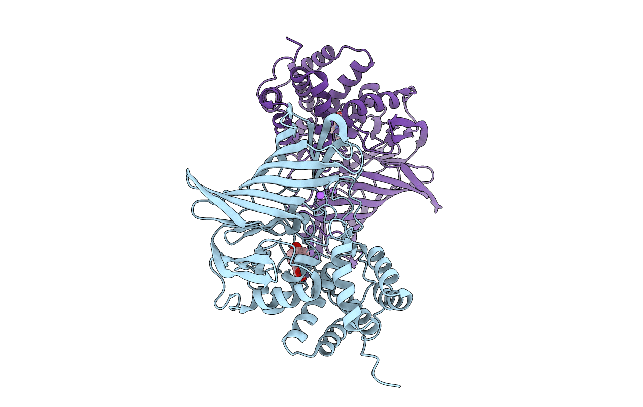
PDB ID:
6A8Z
Keywords:
Title:
Crystal structure of M1 zinc metallopeptidase from Deinococcus radiodurans
Biological Source:
Source Organism:
Host Organism:
Method Details:
Experimental Method:
Resolution:
2.05 Å
R-Value Free:
0.22
R-Value Work:
0.17
R-Value Observed:
0.17
Space Group:
P 1