6A6I image
Deposition Date 2018-06-28
Release Date 2019-02-13
Last Version Date 2024-11-06
Entry Detail
PDB ID:
6A6I
Keywords:
Title:
Crystal structure of the winged-helix domain of Cockayne syndrome group B protein in complex with ubiquitin
Biological Source:
Source Organism:
Homo sapiens (Taxon ID: 9606)
Host Organism:
Method Details:
Experimental Method:
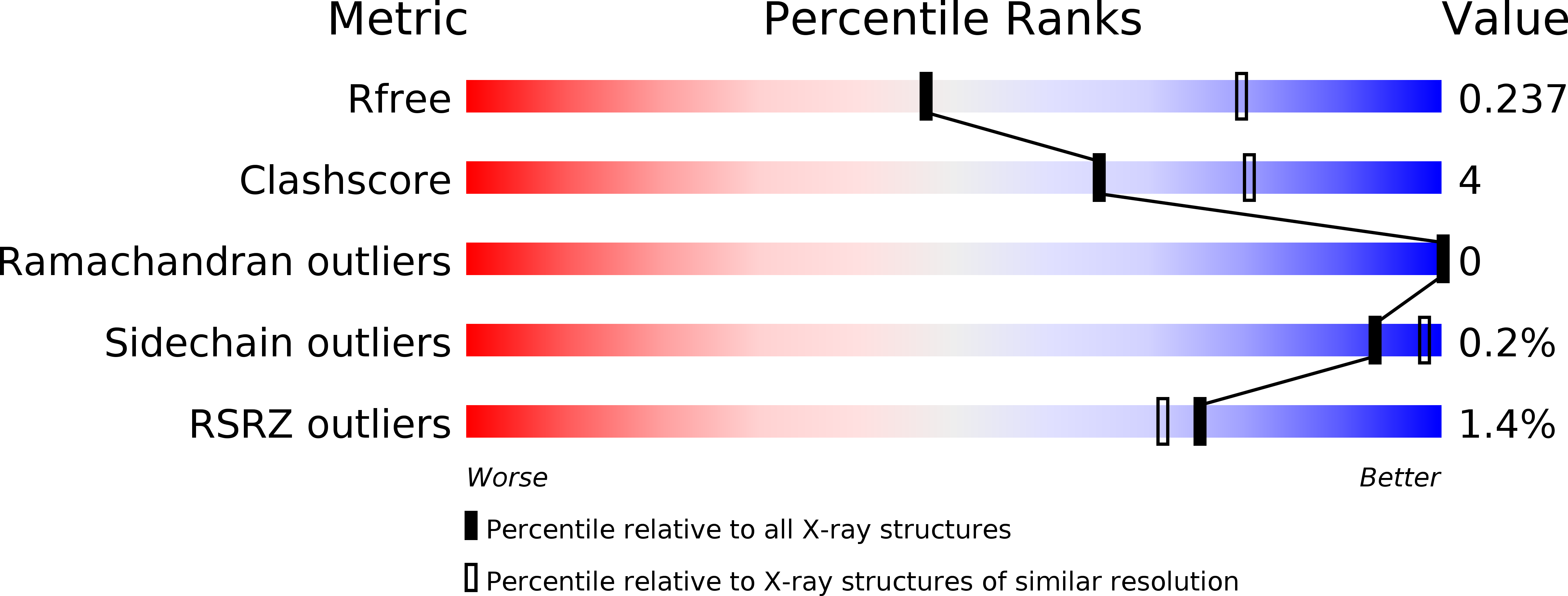
Resolution:
2.60 Å
R-Value Free:
0.23
R-Value Work:
0.17
R-Value Observed:
0.18
Space Group:
P 1 21 1
Macromolecular Entities
Structures with similar UniProt ID
Protein Blast
Polymer Type:polypeptide(L)
Molecule:Excision repair cross-complementing rodent repair deficiency, complementation group 6 variant
Chain IDs:A, C, E, G
Chain Length:98
Number of Molecules:4
Biological Source:Homo sapiens
Structures with similar UniProt ID
Protein Blast
Polymer Type:polypeptide(L)
Molecule:Polyubiquitin-B
Gene (Uniprot):UBB
Mutagens:K48R
Chain IDs:B, D
Chain Length:76
Number of Molecules:2
Biological Source:Homo sapiens
Structures with similar UniProt ID
Protein Blast
Polymer Type:polypeptide(L)
Molecule:Polyubiquitin-B
Gene (Uniprot):UBB
Mutagens:M77D
Chain IDs:F, H
Chain Length:77
Number of Molecules:2
Biological Source:Homo sapiens
Primary Citation
Structural basis of ubiquitin recognition by the winged-helix domain of Cockayne syndrome group B protein.
Nucleic Acids Res. 47 3784 3794 (2019)
PMID: 30753618 DOI: 10.1093/nar/gkz081

Abstact

Cockayne syndrome group B (CSB, also known as ERCC6) protein is involved in many DNA repair processes and essential for transcription-coupled repair (TCR). The central region of CSB has the helicase motif, whereas the C-terminal region contains important regulatory elements for repair of UV- and oxidative stress-induced damages and double-strand breaks (DSBs). A previous study suggested that a small part (∼30 residues) within this region was responsible for binding to ubiquitin (Ub). Here, we show that the Ub-binding of CSB requires a larger part of CSB, which was previously identified as a winged-helix domain (WHD) and is involved in the recruitment of CSB to DSBs. We also present the crystal structure of CSB WHD in complex with Ub. CSB WHD folds as a single globular domain, defining a class of Ub-binding domains (UBDs) different from 23 UBD classes identified so far. The second α-helix and C-terminal extremity of CSB WHD interact with Ub. Together with structure-guided mutational analysis, we identified the residues critical for the binding to Ub. CSB mutants defective in the Ub binding reduced repair of UV-induced damage. This study supports the notion that DSB repair and TCR may be associated with the Ub-binding of CSB.

Legend

Protein

Chemical

Disease

Primary Citation of related structures