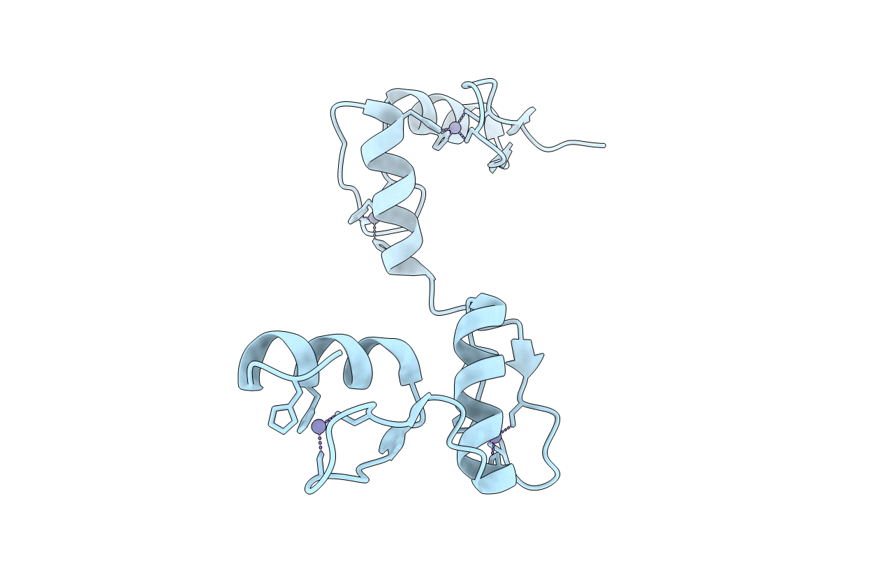
6A59 image
Deposition Date 2018-06-22
Release Date 2019-06-26
Last Version Date 2025-03-12
Entry Detail
PDB ID:
6A59
Title:
Structure of histone demethylase REF6 at 1.8A
Biological Source:
Source Organism:
Host Organism:
Method Details:
Experimental Method:
Resolution:
1.82 Å
R-Value Free:
0.22
R-Value Work:
0.21
R-Value Observed:
0.21
Space Group:
P 41
Macromolecular Entities
Structures with similar UniProt ID
Protein Blast
Polymer Type:polypeptide(L)
Molecule:Lysine-specific demethylase REF6
Gene (Uniprot):REF6
Chain IDs:A
Chain Length:140
Number of Molecules:1
Biological Source:Arabidopsis thaliana
Ligand Molecules
Primary Citation
Crystal structures of REF6 and its complex with DNA reveal diverse recognition mechanisms.
Cell Discov 6 17 17 (2020)
PMID: 32257379 DOI: 10.1038/s41421-020-0150-6

Abstact

Relative of Early Flowing 6 (REF6) is a DNA-sequence-specific H3K27me3/2 demethylase that contains four zinc finger (ZnF) domains and targets several thousand genes in Arabidopsis thaliana. The ZnF domains are essential for binding target genes, but the structural basis remains unclear. Here, we determined crystal structures of the ZnF domains and REF6-DNA complex, revealing a unique REF6-family-specific half-cross-braced ZnF (RCZ) domain and two C2H2-type ZnFs. DNA-binding induces a profound conformational change in the hinge region of REF6. Each REF6 recognizes six bases and DNA methylation reduces the binding affinity. Both the acidic region and basic region are important for the self-association of REF6. The REF6 DNA-binding affinity is determined by the sequence-dependent conformations of DNA and also the cooperativity in different target motifs. The conformational plasticity enables REF6 to function as a global transcriptional regulator that directly binds to many diverse genes, revealing the structural basis for the epigenetic modification recognition.

Legend

Protein

Chemical

Disease

Primary Citation of related structures