
Deposition Date
2018-06-20
Release Date
2018-10-24
Last Version Date
2023-11-22
Entry Detail
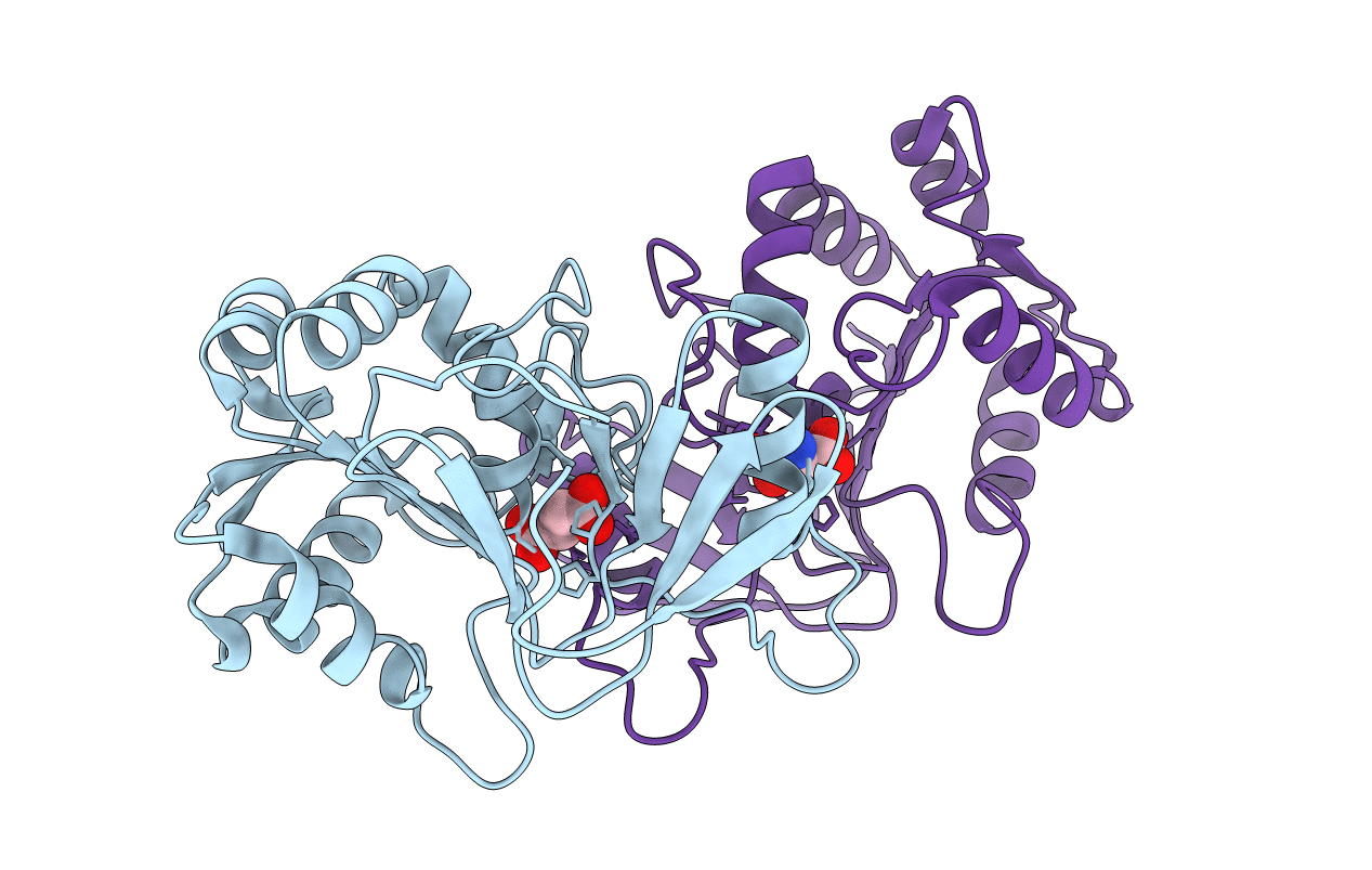
PDB ID:
6A4R
Keywords:
Title:
Crystal structure of aspartate bound peptidase E from Salmonella enterica
Biological Source:
Source Organism(s):
Expression System(s):
Method Details:
Experimental Method:
Resolution:
1.83 Å
R-Value Free:
0.21
R-Value Work:
0.18
R-Value Observed:
0.19
Space Group:
P 1 21 1
