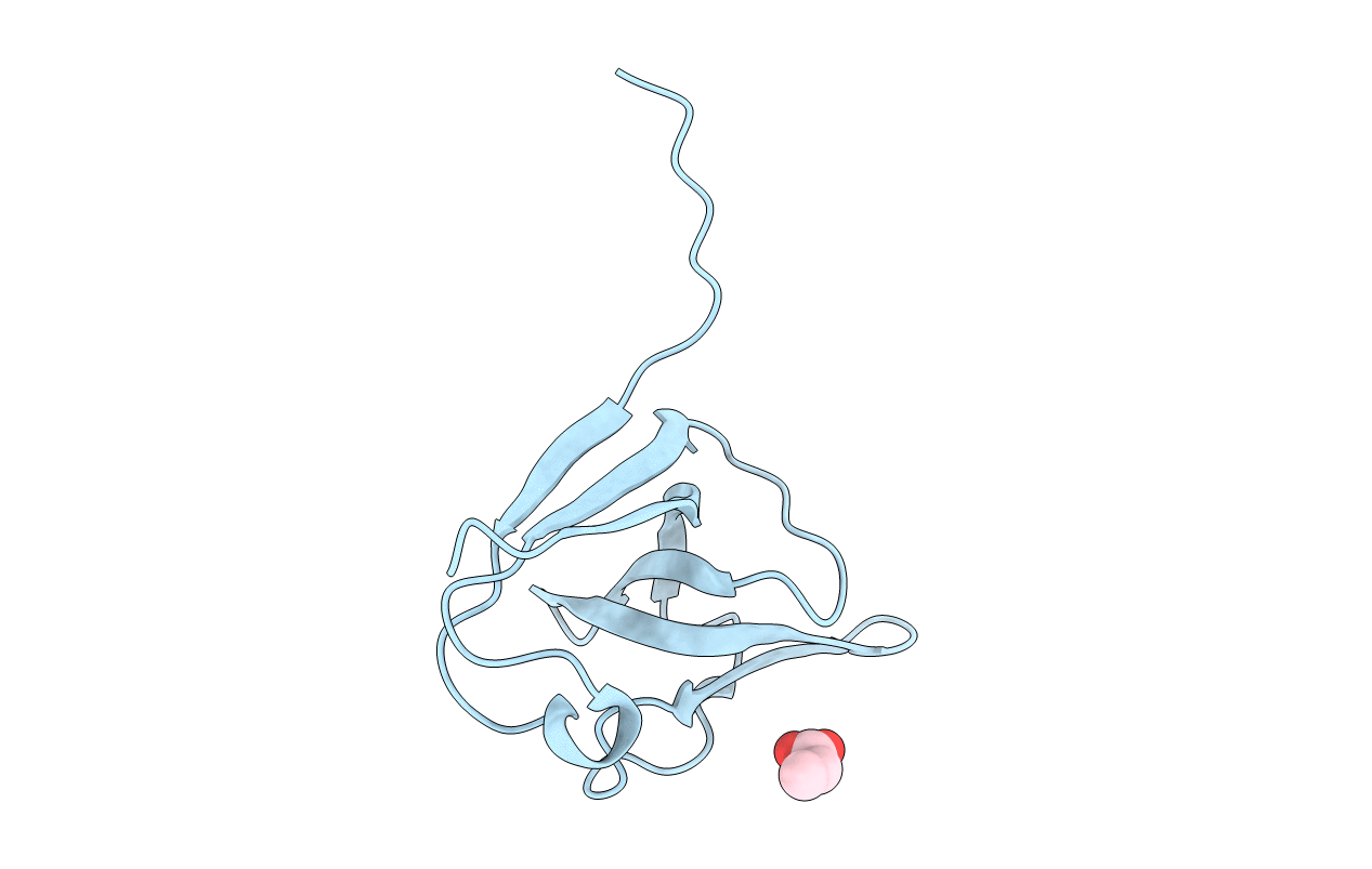
6A2T image
Deposition Date 2018-06-12
Release Date 2019-01-23
Last Version Date 2024-10-30
Entry Detail
PDB ID:
6A2T
Keywords:
Title:
Mycobacterium tuberculosis LexA C-domain K197A
Biological Source:
Source Organism:
Host Organism:
Method Details:
Experimental Method:
Resolution:
1.90 Å
R-Value Free:
0.28
R-Value Work:
0.24
R-Value Observed:
0.24
Space Group:
I 2 2 2
Macromolecular Entities
Structures with similar UniProt ID
Protein Blast
Polymer Type:polypeptide(L)
Molecule:LexA repressor
Gene (Uniprot):lexA
Mutagens:K197A
Chain IDs:A
Chain Length:111
Number of Molecules:1
Biological Source:Mycobacterium tuberculosis
Modified Residue
Compound ID Chain ID Parent Comp ID Details 2D Image
CME A CYS modified residue
Ligand Molecules
Primary Citation
Biochemical characterization of Mycobacterium tuberculosis LexA and structural studies of its C-terminal segment.
Acta Crystallogr D Struct Biol 75 41 55 (2019)
PMID: 30644844 DOI: 10.1107/S2059798318016066

Abstact

LexA is a protein that is involved in the SOS response. The protein from Mycobacterium tuberculosis and its mutants have been biochemically characterized and the structures of their catalytic segments have been determined. The protein is made up of an N-terminal segment, which includes the DNA-binding domain, and a C-terminal segment encompassing much of the catalytic domain. The two segments are defined by a cleavage site. Full-length LexA, the two segments, two point mutants involving changes in the active-site residues (S160A and K197A) and another mutant involving a change at the cleavage site (G126D) were cloned and purified. The wild-type protein autocleaves at basic pH, while the mutants do not. The wild-type and the mutant proteins dimerize and bind DNA with equal facility. The C-terminal segment also dimerizes, and it also shows a tendency to form tetramers. The C-terminal segment readily crystallized. The crystals obtained from attempts involving the full-length protein and its mutants contained only the C-terminal segment including the catalytic core and a few residues preceding it, in a dimeric or tetrameric form, indicating protein cleavage during the long period involved in crystal formation. Modes of tetramerization of the full-length protein similar to those observed for the catalytic core are feasible. A complex of M. tuberculosis LexA and the cognate SOS box could be modeled in which the mutual orientation of the two N-terminal domains differs from that in the Escherichia coli LexA-DNA complex. These results represent the first thorough characterization of M. tuberculosis LexA and provide definitive information on its structure and assembly. They also provide leads for further exploration of this important protein.

Legend

Protein

Chemical

Disease

Primary Citation of related structures