6A0L image
Deposition Date 2018-06-05
Release Date 2018-09-12
Last Version Date 2023-11-22
Entry Detail
PDB ID:
6A0L
Keywords:
Title:
Cyclic alpha-maltosyl-(1-->6)-maltose hydrolase from Arthrobacter globiformis, complex with maltose
Biological Source:
Source Organism:
Host Organism:
Method Details:
Experimental Method:
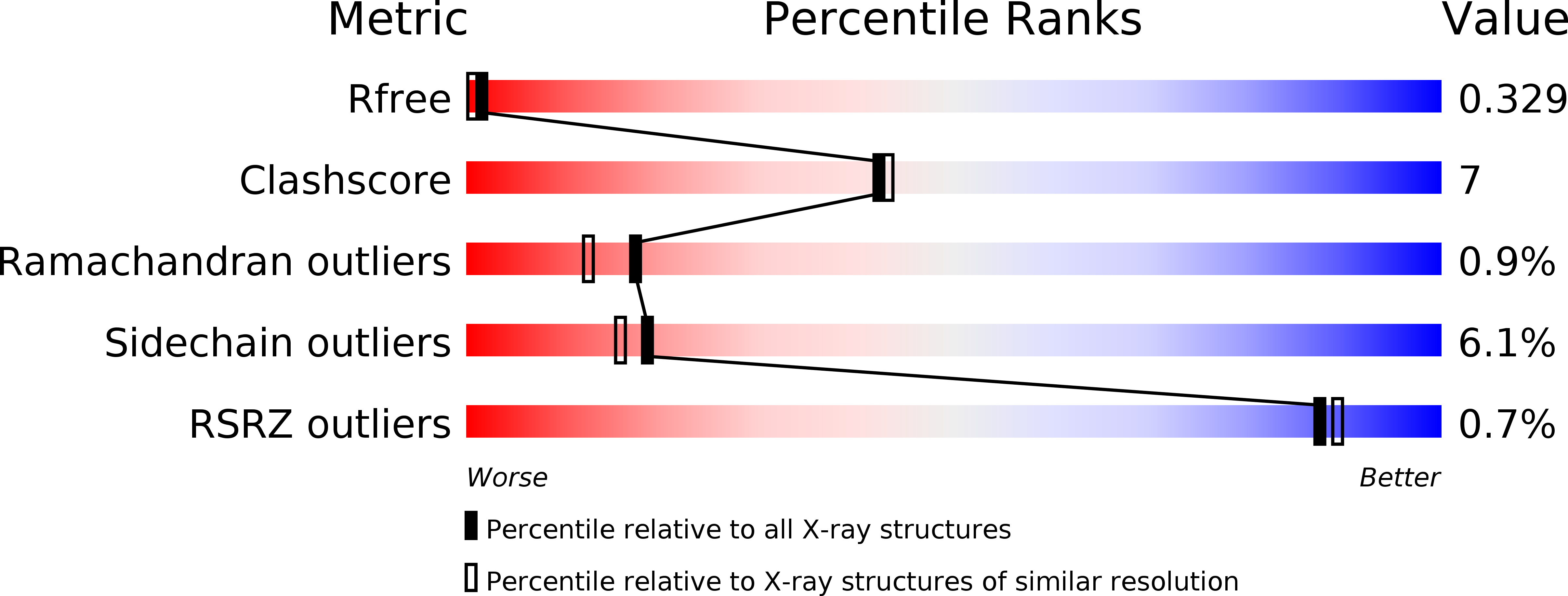
Resolution:
2.10 Å
R-Value Free:
0.32
R-Value Work:
0.25
R-Value Observed:
0.25
Space Group:
C 2 2 21
Macromolecular Entities
Structures with similar UniProt ID
Protein Blast
Polymer Type:polypeptide(L)
Molecule:Cyclic maltosyl-maltose hydrolase
Gene (Uniprot):cmmF
Chain IDs:A
Chain Length:471
Number of Molecules:1
Biological Source:Arthrobacter globiformis
Peptide-like Molecules
PRD_900001
Primary Citation
Structural features of a bacterial cyclic alpha-maltosyl-(1→6)-maltose (CMM) hydrolase critical for CMM recognition and hydrolysis.
J. Biol. Chem. 293 16874 16888 (2018)
PMID: 30181215 DOI: 10.1074/jbc.RA118.004472

Abstact

Cyclic α-maltosyl-(1→6)-maltose (CMM, cyclo-{→6)-α-d-Glcp-(1→4)-α-d-Glcp-(1→6)-α-d-Glcp-(1→4)-α-d-Glcp-(1→})is a cyclic glucotetrasaccharide with alternating α-1,4 and α-1,6 linkages. CMM is composed of two maltose units and is one of the smallest cyclic glucooligosaccharides. Although CMM is resistant to usual amylases, it is efficiently hydrolyzed by CMM hydrolase (CMMase), belonging to subfamily 20 of glycoside hydrolase family 13 (GH13_20). Here, we determined the ligand-free crystal structure of CMMase from the soil-associated bacterium Arthrobacter globiformis and its structures in complex with maltose, panose, and CMM to elucidate the structural basis of substrate recognition by CMMase. The structures disclosed that although the monomer structure consists of three domains commonly adopted by GH13 and other α-amylase-related enzymes, CMMase forms a unique wing-like dimer structure. The complex structure with CMM revealed four specific subsites, namely -3', -2, -1, and +1'. We also observed that the bound CMM molecule adopts a low-energy conformer compared with the X-ray structure of a single CMM crystal, also determined here. Comparison of the CMMase active site with those in other enzymes of the GH13_20 family revealed that three regions forming the wall of the cleft, denoted PYF (Pro-203/Tyr-204/Phe-205), CS (Cys-163/Ser-164), and Y (Tyr-168), are present only in CMMase and are involved in CMM recognition. Combinations of multiple substitutions in these regions markedly decreased the activity toward CMM, indicating that the specificity for this cyclic tetrasaccharide is supported by the entire shape of the pocket. In summary, our work uncovers the mechanistic basis for the highly specific interactions of CMMase with its substrate CMM.

Legend

Protein

Chemical

Disease

Primary Citation of related structures
Feedback Form
Name
Email
Institute
Feedback