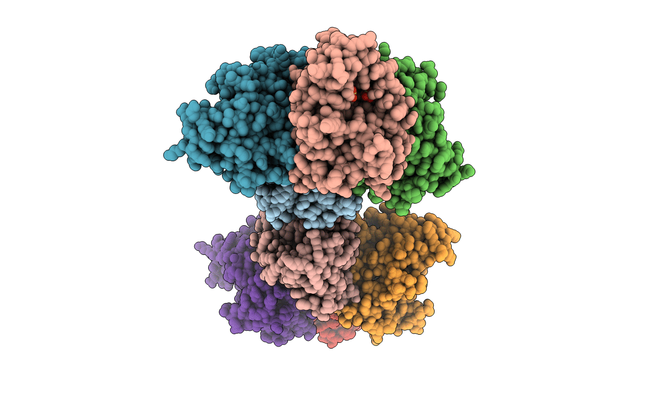
5pgm image
Deposition Date 1998-08-19
Release Date 1999-02-16
Last Version Date 2024-12-25
Entry Detail
PDB ID:
5PGM
Keywords:
Title:
SACCHAROMYCES CEREVISIAE PHOSPHOGLYCERATE MUTASE
Biological Source:
Source Organism:
Method Details:
Experimental Method:
Resolution:
2.12 Å
R-Value Free:
0.22
R-Value Work:
0.19
R-Value Observed:
0.19
Space Group:
P 1 21 1
Macromolecular Entities
Polymer Type:polypeptide(L)
Molecule:PHOSPHOGLYCERATE MUTASE 1
Gene (Uniprot):GPM1
Chain IDs:A (auth: D), B (auth: C), C (auth: A), D (auth: B), E, F, G, H
Chain Length:246
Number of Molecules:8
Biological Source:Saccharomyces cerevisiae
Primary Citation
Sulphate ions observed in the 2.12 A structure of a new crystal form of S. cerevisiae phosphoglycerate mutase provide insights into understanding the catalytic mechanism.
J.Mol.Biol. 286 1507 1517 (1999)
PMID: 10064712 DOI: 10.1006/jmbi.1999.2566

Abstact

The structure of a new crystal form of Saccharomyces cerevisiae phosphoglycerate mutase has been solved and refined to 2.12 A with working and free R-factors of 19.7 and 22.9 %, respectively. Higher-resolution data and greater non-crystallographic symmetry have produced a more accurate protein structure than previously. Prominent among the differences from the previous structure is the presence of two sulphate ions within each active site cleft. The separation of the sulphates suggests that they may occupy the same sites as phospho groups of the bisphosphate ligands of the enzyme. Plausible binding modes for 2,3-bisphosphoglycerate and 1, 3-bisphosphoglycerate are thereby suggested. These results support previous conclusions from mutant studies, highlight interesting new targets for mutagenesis and suggest a possible mechanism of enzyme phosphorylation.

Legend

Protein

Chemical

Disease

Primary Citation of related structures