5i8f image
Deposition Date 2016-02-18
Release Date 2016-05-25
Last Version Date 2024-01-10
Entry Detail
PDB ID:
5I8F
Keywords:
Title:
Crystal structure of St. John's wort Hyp-1 protein in complex with melatonin
Biological Source:
Source Organism:
Method Details:
Experimental Method:
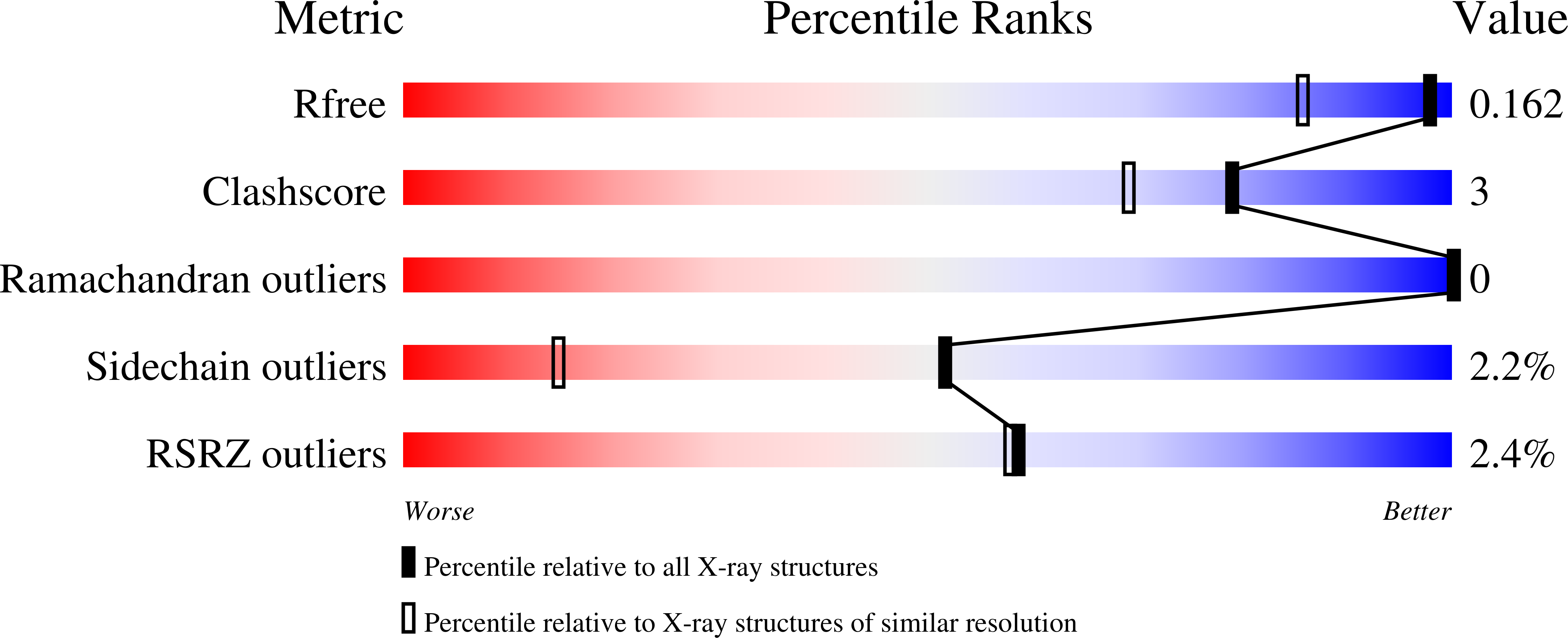
Resolution:
1.30 Å
R-Value Free:
0.15
R-Value Work:
0.12
Space Group:
C 2 2 21
Macromolecular Entities
Structures with similar UniProt ID
Protein Blast
Polymer Type:polypeptide(L)
Molecule:Phenolic oxidative coupling protein
Chain IDs:A
Chain Length:165
Number of Molecules:1
Biological Source:Hypericum perforatum
Primary Citation

Abstact

Hyp-1, a PR-10-fold protein from Hypericum perforatum, was crystallized in complex with melatonin (MEL). The structure confirms the conserved protein fold and the presence of three unusual ligand binding sites, two of which are internal chambers (1,2), while the third one (3) is formed as an invagination of the protein surface. The MEL ligand in site 1 is well defined while that in site 3 seems to be rotating between the side chains of Lys33 and Tyr150 that act as a molecular vise. The patch of electron density in site 2 does not allow unambiguous modeling of a melatonin molecule but suggests a possible presence of its degradation product. This pattern of ligand occupation is reproducible in repeated crystallization/structure determination experiments. Although the binding of melatonin by Hyp-1 does not appear to be very strong (for example, MEL cannot displace the artificial fluorescence probe ANS), it is strong enough to suggest a physiological role of this interaction. For example, trans-zeatin, which is a common ligand of PR-10 proteins, does not overcompete melatonin for binding to Hyp-1 as it does not affect the crystallization process of the Hyp-1/MEL complex, and among a number of potential natural mediators tested, melatonin was the only one to form a crystalline complex with Hyp-1 with the use of standard crystallization screens. Hyp-1 is the second protein in the Protein Data Bank for which melatonin binding has been demonstrated crystallographically, the first one being human quinone reductase.

Legend

Protein

Chemical

Disease

Primary Citation of related structures