Deposition Date
2015-10-18
Release Date
2016-03-30
Last Version Date
2024-11-13
Entry Detail
PDB ID:
5FKP
Keywords:
Title:
Crystal structure of the mouse CD1d in complex with the p99 peptide
Biological Source:
Source Organism(s):
MUS MUSCULUS (Taxon ID: 10090)
Expression System(s):
Method Details:
Experimental Method:
Resolution:
1.80 Å
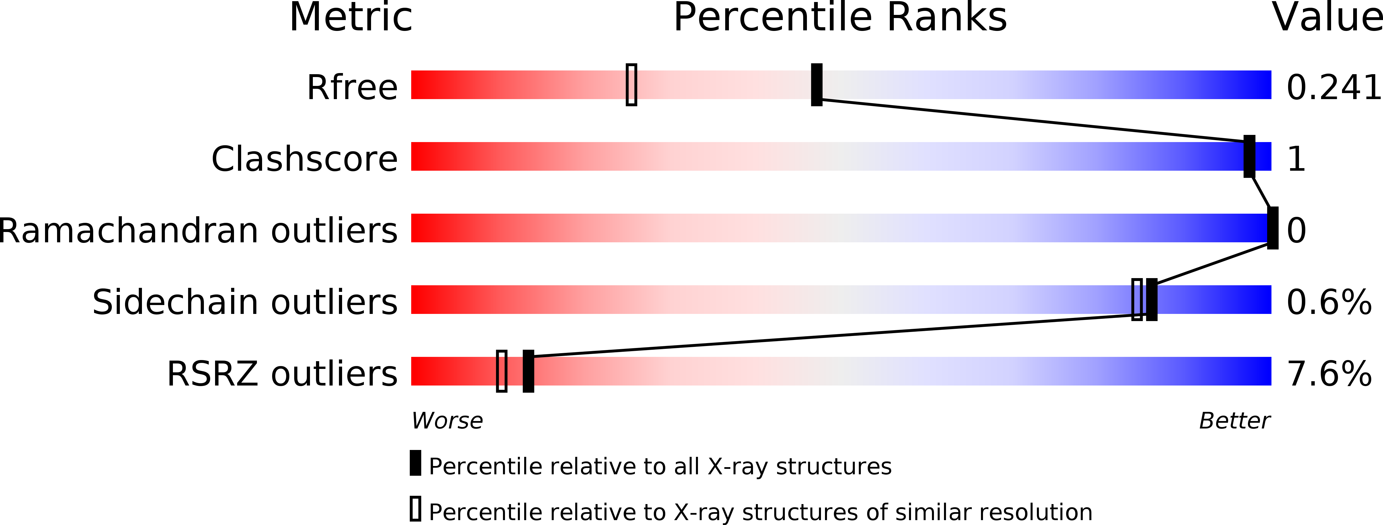
R-Value Free:
0.23
R-Value Work:
0.20
R-Value Observed:
0.20
Space Group:
P 21 21 21