
Deposition Date
2015-12-22
Release Date
2016-07-06
Last Version Date
2024-10-30
Entry Detail
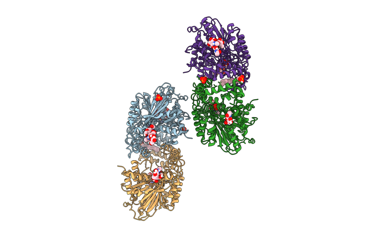
PDB ID:
5FIC
Keywords:
Title:
Open form of murine Acid Sphingomyelinase in presence of lipid
Biological Source:
Source Organism(s):
Mus musculus (Taxon ID: 10090)
Expression System(s):
Method Details:
Experimental Method:
Resolution:
2.80 Å
R-Value Free:
0.23
R-Value Work:
0.18
R-Value Observed:
0.19
Space Group:
P 41
