Deposition Date
2018-04-25
Release Date
2019-10-23
Last Version Date
2024-03-27
Entry Detail
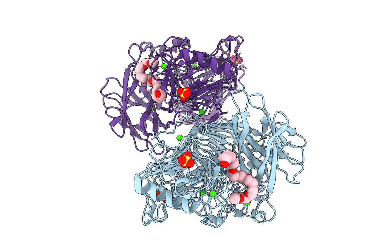
Biological Source:
Source Organism(s):
Bacillus circulans (Taxon ID: 1397)
Expression System(s):
Method Details:
Experimental Method:
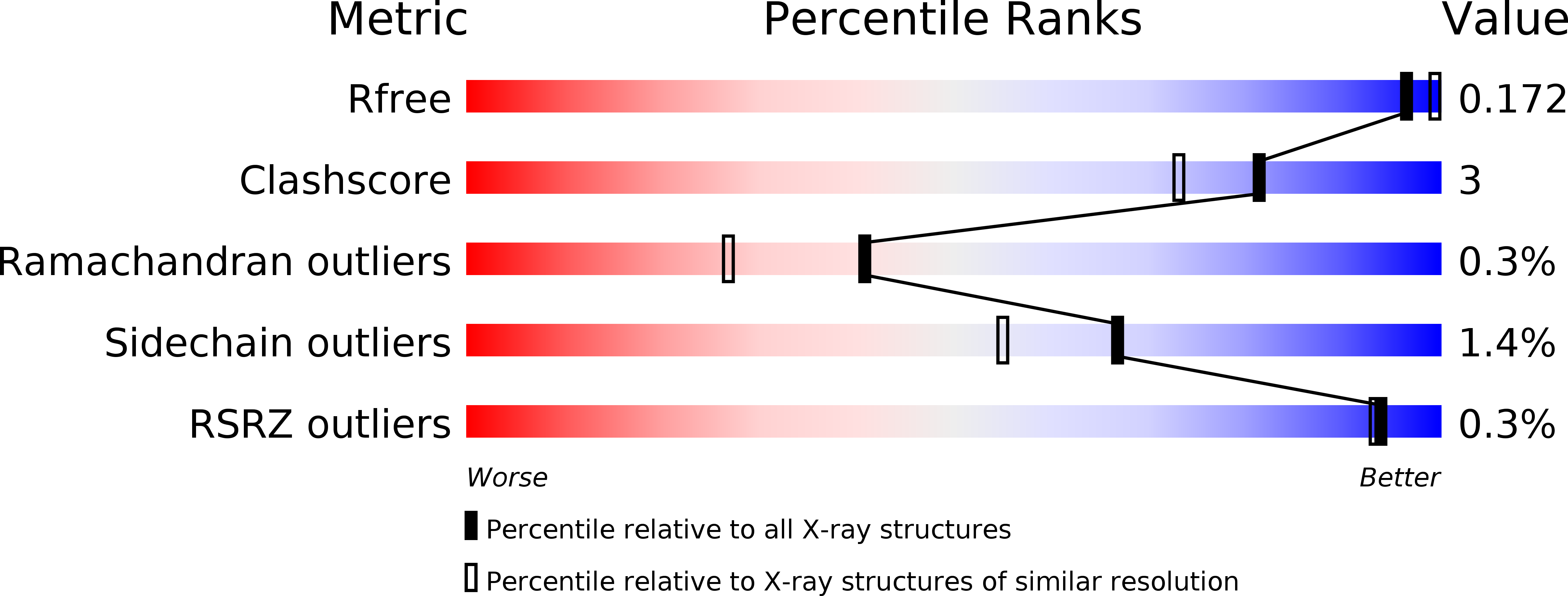
Resolution:
1.83 Å
R-Value Free:
0.17
R-Value Work:
0.14
R-Value Observed:
0.14
Space Group:
P 1 21 1